ARJO Huntleigh Healthcare
Pressure Ulcer Classification Guide
Classification Guide
4 Pages

Preview
Page 1
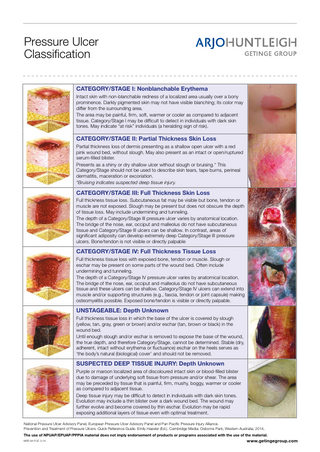
Pressure Ulcer Classification CATEGORY/STAGE I: Nonblanchable Erythema Intact skin with non-blanchable redness of a localized area usually over a bony prominence. Darkly pigmented skin may not have visible blanching; its color may differ from the surrounding area. The area may be painful, firm, soft, warmer or cooler as compared to adjacent tissue. Category/Stage I may be difficult to detect in individuals with dark skin tones. May indicate “at risk” individuals (a heralding sign of risk).
CATEGORY/STAGE II: Partial Thickness Skin Loss Partial thickness loss of dermis presenting as a shallow open ulcer with a red pink wound bed, without slough. May also present as an intact or open/ruptured serum-filled blister. Presents as a shiny or dry shallow ulcer without slough or bruising.* This Category/Stage should not be used to describe skin tears, tape burns, perineal dermatitis, maceration or excoriation. *Bruising indicates suspected deep tissue injury.
CATEGORY/STAGE III: Full Thickness Skin Loss Full thickness tissue loss. Subcutaneous fat may be visible but bone, tendon or muscle are not exposed. Slough may be present but does not obscure the depth of tissue loss. May include undermining and tunneling. The depth of a Category/Stage III pressure ulcer varies by anatomical location. The bridge of the nose, ear, occiput and malleolus do not have subcutaneous tissue and Category/Stage III ulcers can be shallow. In contrast, areas of significant adiposity can develop extremely deep Category/Stage III pressure ulcers. Bone/tendon is not visible or directly palpable
CATEGORY/STAGE IV: Full Thickness Tissue Loss Full thickness tissue loss with exposed bone, tendon or muscle. Slough or eschar may be present on some parts of the wound bed. Often include undermining and tunneling. The depth of a Category/Stage IV pressure ulcer varies by anatomical location. The bridge of the nose, ear, occiput and malleolus do not have subcutaneous tissue and these ulcers can be shallow. Category/Stage IV ulcers can extend into muscle and/or supporting structures (e.g., fascia, tendon or joint capsule) making osteomyelitis possible. Exposed bone/tendon is visible or directly palpable.
UNSTAGEABLE: Depth Unknown Full thickness tissue loss in which the base of the ulcer is covered by slough (yellow, tan, gray, green or brown) and/or eschar (tan, brown or black) in the wound bed. Until enough slough and/or eschar is removed to expose the base of the wound, the true depth, and therefore Category/Stage, cannot be determined. Stable (dry, adherent, intact without erythema or fluctuance) eschar on the heels serves as ‘the body’s natural (biological) cover’ and should not be removed.
SUSPECTED DEEP TISSUE INJURY: Depth Unknown Purple or maroon localized area of discoloured intact skin or blood-filled blister due to damage of underlying soft tissue from pressure and/or shear. The area may be preceded by tissue that is painful, firm, mushy, boggy, warmer or cooler as compared to adjacent tissue. Deep tissue injury may be difficult to detect in individuals with dark skin tones. Evolution may include a thin blister over a dark wound bed. The wound may further evolve and become covered by thin eschar. Evolution may be rapid exposing additional layers of tissue even with optimal treatment. National Pressure Ulcer Advisory Panel, European Pressure Ulcer Advisory Panel and Pan Pacific Pressure Injury Alliance. Prevention and Treatment of Pressure Ulcers: Quick Reference Guide. Emily Haesler (Ed.). Cambridge Media: Osborne Park, Western Australia; 2014. The use of NPUAP/EPUAP/PPPIA material does not imply endorsement of products or programs associated with the use of the material. MRF-84 PUC 3-16
www.getingegroup.com
Weight conversion Stones (St)
Kilograms
Stones (St)
Kilograms
1
6.4
30
190.7
2
12.7
31
196.8
3
19.1
32
200.0
4
25.4
33
209.5
5
31.8
34
216.0
6
38.1
35
222.5
7
44.5
36
228.6
8
50.8
37
235.0
9
57.2
38
241.3
10
63.5
39
247.6
11
69.9
40
254.0
12
76.3
41
260.3
13
82.6
42
266.7
14
89.0
43
273.0
15
95.3
44
279.4
16
101.7
45
285.8
17
108.0
46
292.1
18
114.4
47
298.5
19
120.8
48
304.8
20
120.8
49
311.2
21
127.0
50
317.5
22
133.5
51
323.9
23
139.8
52
330.2
24
146.2
53
336.6
25
152.5
54
343.0
26
158.9
55
349.3
27
165.2
56
355.6
28
171.6
57
362.0
29
184.3
58
363.3
Foam Support SurfaceSUPPORT Testing SURFACE TESTING FOAM Why test foam mattress replacements? • If continually subjected to load, foam support surfaces can suffer a reduction in performance over time due to compression • If the cover material is compromised in any way the core is vulnerable to the ingress of fluids and poses an infection control risk • Whilst an effective turning regime and regular inspections will help prevent compression, it is vital that regular tests are carried out and recorded in order to verify the condition of the mattress and ensure patients are properly supported at all times NOTE: To facilitate effective testing the date that each mattress is installed must be recorded clearly on the cover. This information is a guide only, the responsibility for the final decision regarding the condition of the mattress lies with the auditors. Whenever possible, a Tissue Viability Nurse and an Infection Control Nurse should be present during the audit.
How is the test performed? 1. Record Mattress Details • Note the age of the mattress • Manufacturer
4
• Rental / trial or purchased equipment
3
2. Cover material integrity and condition • Unzip the cover, check for staining, moisture and odour both inside and out
• Any PVC covers should be removed in accordance with the Kings Fund recommendation, 1998
Fist test to assess compression The test is designed to assess whether the foam core has compressed over time, increasing the possibility of the patient bottoming out. With the cover in place and fully zipped: • Join hands together, with straight arms forming a fist (see figure 1) • With the mattress at hip level, push down using full body weight • Repeat the test in the areas indicated in figure 2 • If the base of the mattress can be felt through the foam (bottoming out) the mattress should be removed for further
MRF-69
Midline
7
10cm
• Check for delamination and splitting including zips and seams
3. Foam Core testing
5
1
2
The cover material should be examined in the following way:
If damage or staining on the INSIDE of the cover is found, the entire mattress (core and cover) should be condemned immediately. If staining to the outside of the cover is found, only the cover must be replaced (after careful checking of the foam core, see below)
6
65cm
20cm
Figure 1
Midline
Figure 2
investigation NOTE: If a patient has been lying on the mattress prior to the test, at least three minutes recovery time is required before the torso section can be tested. Foam Core Contamination Fully examine the foam for any staining, splitting, odour or dampness. If ANY of these are found the mattress should be condemned immediately to avoid an infection control risk.
30° Tilt 30° TILT Recumbent
Semi-recumbent
1 1
2
The patient should lie in the centre of the bed with adequate pillows to support the head and neck.
3
Add a pillow to give support to the lumbar region and shoulder. This ‘tilts’ the patient by 30° and lifts the sacrum clear of the mattress. Check with your hand that the sacrum clears the mattress.
The patient’s lower back should be supported with pillows. The lower pillow can be bunched or folded if necessary.
2
4 Another pillow is placed beneath the others, with the corner carefully positioned under the buttock to ‘tilt’ the body and lift clear the ischial tuberosities and sacrum.
3 Support both legs by placing pillows lengthways, ensuring that heels are kept lifted from the bed surface.
5
6 The legs must be supported in the same manner as fig 3 and 4 of the Recumbent position. It is important that the heels are clear and the feet are in the correct position.
4
Patient in full recumbent '30° tilt’ position.
An additional pillow can be placed as shown, to prevent 'foot drop'.
REMEMBER 1. Always explain the whole procedure to the patient, prior to repositioning and throughout, to reassure them. 2. Confirm that the patient is comfortable and check their position frequently. 3. The '30° tilt’ is useful for patient comfort and pressure reduction over high risk areas. It should be used as an aid to, not in place of, an appropriate pressure reducing support surface/mattress.
Patient in complete semi-recumbent '30° tilt’ position.
REMEMBER 1. The use of electric profiling beds will assist in the management of patient positioning.
Reference Preston KW (1989). Positioning for comfort and pressure relief: The 30 degree alternative. Care -Science and Practice; 6(4): 116-119.
MRF-71