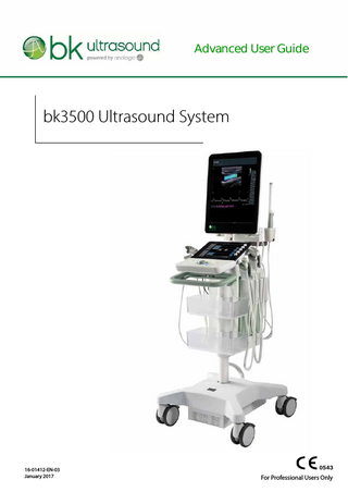
Advanced User Guide
218 Pages

Preview
Page 1
Advanced User Guide
bk3500 Ultrasound System
16-01412-EN-03 January 2017
For Professional Users Only
LEGAL MANUFACTURER BK Medical Aps Mileparken 34 DK-2730 Herlev Denmark Tel.:+45 4452 8100/Fax:+45 4452 8199 www.bkultrasound.com Email: [email protected]
The serial number label on a BK Medical product contains information about the year of manufacture.
BK Medical Customer Satisfaction
Input from our customers helps us improve our products and services. As part of our customer satisfaction program, we contact a sample of our customers a few months after they receive their orders. If you receive an email message from us asking for your feedback, we hope you will be willing to answer some questions about your experience buying and using our products. Your opinions are important to us. You are of course always welcome to contact us via your BK Medical representative or by contacting us directly. If you have comments about the user documentation, please write to us at the email address above. We would like to hear from you. •
•
•
Scanner Software NOT FAULT TOLERANT. THE SOFTWARE IS NOT FAULT TOLERANT. BK Medical HAS INDEPENDENTLY DETERMINED HOW TO USE THE SOFTWARE IN THE DEVICE, AND MS HAS RELIED UPON BK Medical TO CONDUCT SUFFICIENT TESTING TO DETERMINE THAT THE SOFTWARE IS SUITABLE FOR USE. EXPORT RESTRICTIONS. You acknowledge that Windows 8 Embedded is of US-origin. You agree to comply with all applicable international and national laws that apply to Windows 8 Embedded, including the U.S. Export Administration Regulations, as well as end-user, end-use and country destination restrictions issued by U.S. and other governments. For additional information on exporting Windows 8 Embedded, see http:// www.microsoft.com/exporting/ The bk3500 Ultrasound System is closed. Any modification of or installation of software to the system may compromise safety and function of the system. Any modification of or installation of software without written permission fromBK Medical will immediately void any warranty supplied by BK Medical. Such changes will also void any service contract and result in charges to the customer for restoration of the original bk3500 Ultrasound System.
Trademarks: DICOM® is the registered trademark of the National Electrical Manufacturers Association for its standards publications relating to digital communications of medical information. FireWire™ is a trademark of Apple Computer, Inc. Microsoft® and Windows® are registered trademarks of Microsoft Corporation in the United States and other countries.
bk3500 = Ref. Type 2300 © 2017 BK Medical Information in this document may be subject to change without notice.
Contents Chapter 1
Before You Begin... 11
Chapter 2
Getting Started... 13 The bk3500 System... 13 Before You Start... 14 Turning System On and Off... 14 Connecting Transducers... 15 Barcode Reader... 15 Control Panel... 16 Quick Access... 17 Quick Exam Start-Up... 17 Starting an Exam Using the Touch Screen Buttons... 23 Monitor and Touch Screen Display... 26 Documents... 28 Measurements and Image Data... 28
Chapter 3
Controls on the UI... 29
Chapter 4
Working with the Image... 33 Freezing the Image... 33 Partial Freeze and the Update Key... 33 Split Screen... 33 Simultaneous Imaging... 34 Labels and Bodymarks... 35 Labels... 35 Bodymarks... 36 Cine... 38 Storing a Video Clip... 38 Using Cine... 39 Using Cine in M-Mode or Doppler Mode... 40 Video Display... 40
Chapter 5
Making Measurements... 41 Measurements and Calculations... 41 Making a Measurement – General Procedure... 44 B-Mode and Color Mode Measuring Tools... 46 Doppler Mode Measuring Tools... 49
Chapter 6
Documentation... 51 What are Documents?... 51 HIPAA Compliance... 51 Saving Documents – Capturing Images... 51 Capturing Images... 51 Saving Reports... 51 Local Patient Archiving System... 52
3
Reviewing Documents... 52 The Document Browser... 52 Viewing Exported Documents on the System... 53 Viewing Exported Documents on an External Computer... 54 Exporting Data... 55 HIPAA Compliance and Exporting Data... 55 Copying to a USB Storage Device... 55 Archiving to a Network Drive... 56 Using USB Storage Devices... 56 Using a Network Drive... 56 The Archive Window (Examination List and Patient Information)... 56 Patient Information... 57 Examination List... 57 Deleting Documents or Exams from the System... 59 Starting a New Examination from the Examination List... 60 Hard Disk Quota... 61 Worksheets... 61 Reports... 64 Creating a Report... 64 Editing a Report... 65 Printing a Report... 65 Saving a Report to the Local Patient Archiving System... 66 Printing Documents or Images on the Monitor... 66 Printing Thumbnail Images... 66 Printing Images Displayed on the Monitor... 66
Chapter 7
Imaging Modes... 67 Imaging Modes... 67 Adjusting the Thermal Index Limit... 67 B-Mode... 68 Focus... 68 Gain... 69 Auto Gain... 70 Sector... 70 Size... 70 Zoom... 71 Depth... 71 Gray Scales... 71 Tissue Harmonic Imaging (True Echo Harmonics – TEH)... 71 X-Shine... 72 Activate X-Shine Imaging... 74 Combination Modes... 74 Color Mode and Power Mode... 75 Color Submodes... 75 Color Coding of Flow... 75 Color Box... 75 Color Scales... 76 Vector Flow Imaging (VFI)... 76 Using VFI... 78
4
Streamlined VFI Workflow... 79 Assisted Doppler Gate Placement... 80 Angle Correction... 80 Assisted Doppler Steering... 80 Inverting the Doppler Spectrum... 80 Selecting the Appropriate Scale/PRF... 80 Assisted Volume Flow Rate Estimation... 81 Doppler Mode – Spectral Doppler... 82 Turning Doppler Mode On or Off... 82 Adjusting the Doppler Mode Image... 83 Doppler Indicator... 83 Duplex and Triplex... 84 Independent D-Mode/C-Mode Steering... 84 Auto... 84 Doppler Trace (Automatic Curve Tracing)... 84 M-Mode... 85 The M-Mode Image... 85 M-Mode Line... 86 M-Mode Image Ruler... 86
Chapter 8
Continuous Wave Doppler Mode... 89 Overview... 89 Adjusting the Thermal Index Limit... 89 Adjusting the Thermal Index Limit... 89 Turning CW Doppler Mode On or Off... 90 CW Doppler Line... 91 CW Controls on the Touch Keyboard... 92 Audio Volume... 92 Adjusting the Doppler Mode Image... 92 Doppler Trace (Automatic Curve Tracing)... 92 Auto... 93 Gain... 93 Scale... 93 Wall Filter... 93 Invert... 94 Baseline... 94 Sweep Speed... 94
Chapter 9
Applications... 95 Before You Begin... 95 If You Perform a Puncture Procedure... 95 What Is an Application?... 95 Presets... 96 Measurements... 97 Doppler Measurements... 98 Stenosis... 99 VF (Volume Flow)... 100 TAM (Time Average Mean) and TAMX (Time Average Max)... 100 RI and PI (Resistance Index and Pulsatility Index)... 101
5
Real-Time Measurements... 102 Carotid Velocities... 103 Calculations... 103 Using the OB and Gyn Applications... 103 Gestational Age and Expected Date of Confinement... 104 Patient Setup... 104 Making Measurements... 104 Calculation Methods... 104 Obstetrics Reports... 105 Using the Cardiac Application... 105 Measurements and Calculations... 105 Basic Cardiac Measurements... 105 LVV (Left Ventricular Volume)... 106 LAV (Left Atrium Volume)... 109 RAV (Right Atrium Volume)... 110 Doppler Measurements... 110 HR (Heart Rate)... 112 M-Mode Measurements... 112 FATE (Focus Assessed Transthoracic Echocardiography)... 113 FATE Measurements... 113 Where to Find More Information... 115
Chapter 10
DICOM...117 DICOM on the System... 117 New Patient Information from a DICOM Worklist... 117 Saving or Printing to a DICOM Network... 117 Filenames of Documents Exported in DICOM Format... 117 Archiving to a PACS... 117 Reports... 118 Deleting a Document... 119 Discontinuing an Examination with an MPPS Server... 119
Appendix A
6
Setting Up and Customizing Your System... 121 Setup and Customize... 121 General Tab... 122 Layout... 122 Thermal Index Type... 123 Other Settings... 123 User Preferences... 123 Measurement Settings Tab... 124 Author/Method Settings... 125 Result Settings... 127 Cardiac Settings... 128 Worksheets Tab... 129 Worksheet Export... 129 Worksheet Import... 131 Worksheet Assignments... 133 Reset Worksheet to Factory... 135 The System Tab... 136
Remote Support... 136 Service Mode... 137 Wi-Fi... 137 Operator List... 139 Customize... 142 Doppler and M-Mode Monitor Layout... 142 System Setup... 143 General Setup... 143 Clip Storage and Cine Setup... 145 Printer Setup... 146 Network Archiving... 148 Version Information... 149 Video I/O Setup... 150 Battery Support Setup... 151 Miscellaneous System Setup... 152 Measurements... 154 Curves... 154 Miscellaneous Measurement Setup... 157 Licenses... 158 Importing and Exporting System Configurations... 159 Importing or Exporting Presets... 159 Use Pro Packages (Applications)... 161 DICOM Setup... 162 Advanced... 162 About... 163 Labels Tab... 164 Word Library Management... 164 Assign Word Libraries... 169 Import Word Libraries... 170 Export Word Libraries... 171 Reset Word Libraries to Factory... 172 Appendix B
Glossary... 173
Appendix C
Measurement Abbreviations... 179 Generic Measurements... 179 Generic... 179 Generic Doppler... 179 Generic M-Mode... 181 Abdomen... 181 Abdomen (B-Mode)... 181 Aorta/IVC (B-Mode)... 181 Right Kidney (B-Mode)... 182 Left Kidney (B-Mode)... 183 Generic (B-Mode and M-Mode)... 183 Abdomen (Doppler)... 183 Aorta/IVC (Doppler)... 183 Right Renal (Doppler)... 184 Left Renal (Doppler)... 184
7
Generic (Doppler)... 184 Carotid... 184 Right Carotid - Doppler... 184 Left Carotid - Doppler... 185 Generic... 185 OB... 186 OB... 186 Advanced OB... 188 AFI... 188 Early OB... 188 Estimated Fetal Weight... 190 Maternal Doppler... 190 Fetal Doppler... 191 Generic... 191 Early OB... 192 Early OB... 192 Right Ovary... 193 Rt Ovary Doppler... 193 Left Ovary... 193 Lt Ovary Doppler... 193 Uterus... 194 Uterus Doppler... 194 OB... 195 Fetal Doppler... 196 Generic... 196 Gyne... 196 Uterus (B-Mode)... 196 Right Ovary (B-Mode)... 196 Left Ovary (B-Mode)... 197 Bladder (B-Mode)... 197 Generic (B-mode and M-Mode)... 197 Uterus (Doppler)... 197 Rt Ovary (Doppler)... 198 Lt Ovary (Doppler)... 198 Generic (Doppler)... 198 Vascular... 198 Vascular (B-Mode)... 198 Generic (B-Mode, M-Mode and Doppler)... 198 Cardiac... 198 LV / LA (B-Mode)... 198 RV / RA (B-Mode)... 200 Ao / IVC (B-Mode)... 200 LV / RV (M-Mode)... 200 LA /AO / IVC M-Mode... 201 FATE (M-Mode)... 201 Valves (M-Mode)... 201 LV / RV (Doppler)... 202 AV / MV ( Doppler)... 202 PV / TV (Doppler)... 202
8
Generic Doppler... 203 Cardiac Advanced... 205 LV / LA (B-Mode)... 205 RV / RA (B-Mode)... 207 Ao / IVC (B-Mode)... 207 LV / RV (M-Mode)... 207 ... 208 FATE (M-Mode)... 208 Valves (M-Mode)... 208 LV / RV (Doppler... 209 AV / MV ( Doppler)... 209 PV / TV (Doppler)... 210 Generic (Doppler)... 210 Renal... 210 Right Kidney (B-Mode)... 210 Left Kidney (B-Mode)... 210 Aorta/IVC (B-Mode)... 211 Bladder (B-Mode)... 211 Generic B Mode... 211 Generic M-Mode... 212 Right Renal ( Doppler)... 212 Left Renal (Doppler)... 212 Aorta/IVC (Doppler)... 212 Generic Doppler... 212 Index
213
9
Chapter 1 Before You Begin This is the advanced user guide for the bk3500 Ultrasound System. The bk3500 User Guide includes an overview of all the documentation available for the system, including different user guides. NOTE: You must read the Safety chapter in the bk3500 User Guide before working
with the system. This guide takes you deeper into the functionality and potential of the bk3500 Ultrasound System. NOTE: Some of the functionality and options described in this guide may not be
available with your version of the system. Questions About the System
Where to Find the Answers
How do I get started and what are the various parts of the touch screen and monitor displays?
“Getting Started” on page 13
Is there an alphabetical list of all the controls on the system?
“Controls on the UI” on page 29
How do you make measurements and calculations for an image, and what measurement tools are available?
“Making Measurements” on page 41
How do you manage the images, clips and reports that are made on the system?
“Documentation” on page 51
What imaging modes are available on the bk3500?
“Imaging Modes” on page 67
What is an examination type, and how does it help with imaging?
“Applications” on page 95
How does DICOM® work with the bk3500?
“DICOM” on page 117
What do various abbreviations mean?
“Glossary” on page 173
bk3500 Advanced User Guide (16-01412-EN-03)
Before You Begin
11
12 Chapter 1
January 2017
bk3500 Advanced User Guide (16-01412-EN-03)
Chapter 2 Getting Started The bk3500 System
Monitor
Endotransducer holder Touch screen Control panel
Transducer holders
Front handle with release handles
Transducer sockets Scan engine
Baskets for accessories
Lockable wheels Battery display Battery cover
Battery compartment
bk3500 Advanced User Guide (16-01412-EN-03)
Getting Started
13
Before You Start Before you turn on the system, make sure that the installation has been approved by a qualified electrician or by hospital safety personnel. Read the battery support warnings (warnings with BS numbers) in the Battery Support System section of the bk3500 User Guide.
Turning System On and Off When you turn the system on or off, you must give the system enough time to save and recover open files and unsaved data. Otherwise, a serious system failure may occur that requires technical support. Make sure the battery is charged. (If it is not, plug in the imaging system to use it or to charge the battery.)
Figure 2-1. The power button on the scan engine.
To turn on: Press the power button once, then wait until startup screen disappears. If the battery is empty, it is not necessary to turn off the imaging system. Plug the system into a power outlet to recharge the battery while you run on power from the mains power supply. To turn off: Make sure system is running. Press the power button once. Caution BS-c2 Never shut down a system with a battery module simply by unplugging it from the wall. To preserve battery power, shut down the system properly.
14 Chapter 2
January 2017
bk3500 Advanced User Guide (16-01412-EN-03)
Connecting Transducers
Figure 2-2. Transducer plugs and sockets.
To connect: 1 Insert transducer plug into socket with locking lever to the right. 2 Turn locking lever on socket to the left. To disconnect: 1 Freeze image. 2 Turn locking lever on socket to the right. 3 Remove plug from socket. NOTE: If more than one transducer is connected, select a different transducer before you disconnect. Otherwise, the following message will be displayed on the touch screen:
Figure 2-3. Message if the active transducer is disconnected.
Barcode Reader To enter Patient Information with the barcode reader: 1 Tap the touch screen Patient button, or press Q to open the patient dialog when exam is not running.
bk3500 Advanced User Guide (16-01412-EN-03)
Getting Started
15
2 3
With the cursor in the Patient ID field, scan the relevant patient barcode with the barcode reader. Continue entering the patient/exam data as required.
NOTE: Fields that will accept data entry via the keyboard will also accept data scanned with the barcode reader. Simply ensure that the cursor is located in the required field then scan the relevant barcode. WARNING SR-w2 To avoid personal injury, connecting/disconnecting the barcode reader and/or printer must be carried out only by BK personnel or authorized representatives.
Control Panel
Figure 2-4. The control panel and touch screen.
Icon
16 Chapter 2
System Control
Functionality
Trackball
Positions the mouse cursor, measurement cursor and label.
January 2017
bk3500 Advanced User Guide (16-01412-EN-03)
Icon
System Control
Functionality
QUICK ACCESS button
Opens the quick exam start-up workflow. When the exam has started, the Q button works as an Auto button which will automatically optimize the image settings.
Live image: Stores a prospective video clip.
•
2 Button
Frozen image: Stores a retrospective video clip.
SELECT Button
Provides a wide variety of functions depending on the imaging state, for example toggles between moving/resizing the color box and selects/sets measurements, labels, etc.
UPDATE Button
Provides a wide variety of functions depending on the imaging state, for example toggles between image views and image mode and rotates the transducer on the bodymark icon. Stores the current image.
1 Button
FREEZE Button
Freezes/unfreezes live imaging. A snowflake icon is displayed on the monitor when the image is frozen.
Touch Screen Dials
Five dials that control touch screen options, which change depending on the imaging mode/state. Once the touch screen option is tapped, turn the related dial to make the relevant adjustments.
Touch Screen
Displays selectable options. Touch screen buttons may change depending on the chosen imaging mode/state or action.
Quick Access The Q button provides the following basic functions: •
Quick exam start-up
Quick Exam Start-Up Once the Q button is selected, users can navigate through the Quick Exam Startup using the touch screen: 1 Enter Patient Information.
bk3500 Advanced User Guide (16-01412-EN-03)
Getting Started
17
2 3 4 5
18 Chapter 2
Select Transducer. Select Application (Exam type). Select Imaging Preset. Begin the exam.
January 2017
bk3500 Advanced User Guide (16-01412-EN-03)
For the Quick Exam Start-Up: 1 Press the control panel Q button. 2 Enter Patient information. The Patient ID is filled in automatically with a timestamp, but you can change this to a relevant ID or use a barcode reader. See “Barcode Reader” on page 15.
Figure 2-5. Patient window.
3
Swipe the screen from right to left to enter additional patient information.
Figure 2-6. Second screen in Patient window.
bk3500 Advanced User Guide (16-01412-EN-03)
Getting Started
19
4
Tap the Exam Info button to add specific information relevant for the exam, and tap Next.
Figure 2-7. Exam Info window.
5
Select Transducer (in this case 6C2 is selected).
Figure 2-8. List of available transducers (those that are plugged in).
20 Chapter 2
January 2017
bk3500 Advanced User Guide (16-01412-EN-03)
6
Select Application (the exam type you intend to perform). The applications available depend on the selected transducer (in this case 6C2).
Figure 2-9. List of available applications.
7
Select imaging Preset: The imaging presets available are dependent on the transducer and the application (exam type) selected.
Figure 2-10. Available presets.
bk3500 Advanced User Guide (16-01412-EN-03)
Getting Started
21