BK Medical
bkFusion Setting Up - Transrectal Getting Started Guide Ref 2300 Oct 2018
Getting Started Guide
26 Pages

Preview
Page 1
Getting Started Guide
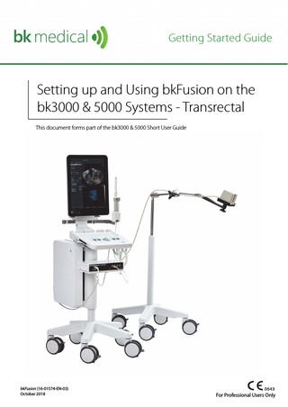
Setting up and Using bkFusion on the bk3000 & 5000 Systems - Transrectal This document forms part of the bk3000 & 5000 Short User Guide
bkFusion (16-01574-EN-03) October 2018
For Professional Users Only
Table of Contents General Information...5 bkFusion Hardware Configuration...5 Configuring bkFusion Hardware...6 MRI Data Transfer from USB/CD, MIMcloud, Remote Patient List & PACS...7 To Review a BXplan...9 To Contour/Re-contour MRI Slices... 11 Predictive Fusion... 15 To Start the Procedure... 17 To Biopsy with bkFusion... 18 To Make Measurements... 21 To Save Biopsy Results and Create a Report... 22 To Save a Report to a USB device... 23 To Save Structured Reports... 23 To Save DICOM Images... 23
bkFusion (16-01574-EN-03)
3
4
October 2018
bkFusion (16-01574-EN-03)
General Information This user guide forms part of the bk3000 & bk5000 user guide. Please refer to the bk3000 & bk5000 user guide for safety information regarding bkFusion. Before using the equipment, please make yourself familiar with the information in the accompanying user information documents. Some documents are printed. Make sure that you also read the transducer user guide and specifications for each transducer that you use.
bkFusion Hardware Configuration The following image shows the hardware configuration of bkFusion:
EM Sensor and E14C4t Transducer The Sensor attaches via the Sensor Clamp to the Transducer. The Transducer transmits ultrasound data to the System. The position of the Sensor is detected by the EM Transmitter.
EM Transmitter (mounted on stand) The EM Transmitter detects the position of the Sensor electromagnetically, and transmits the data to the EM Control Unit.
bk3000 Ultrasound System with bkFusion Software The System receives ultrasound data from the Transducer, and Sensor positioning data from the EM Control Unit. EM Control Unit The EM Control Unit receives the position of the sensor from the EM Transmitter, and transmits the position of the Sensor to the ultrasound system.
Figure 2-1. bkFusion hardware configuration
bkFusion (16-01574-EN-03)
5
Configuring bkFusion Hardware bkFusion is compatible with the USB foot switch, which can be set as ‘mouse left-click’. See the bk3000 & bk5000 Advanced User Guide for foot switch configuration information. 1. Connect the EM Control Unit to the bk3000 system using the USB cable (USB ports are located in the rear of the CPU). Connect the power cable, then switch the unit ON.
2. Connect the EM Transmitter to the EM Control Unit: Insert the Transmitter cable into the Transmitter socket until an audible ‘click’ is heard.
3. Connect the Sensor to the EM Control Unit: Insert the Sensor cable into socket 1 until an audible ‘click’ is heard.
E14C4T (9018)
E13C2 (9029) Open the Sensor Clamp and push the Sensor into the channel, as indicated. The small indentation on the tip of the Sensor must be facing the groove in the Sensor Clamp channel.
4. Push the Sensor into the Sensor Clamp channel. The small indentation on the tip of the Sensor must be facing the groove in the Sensor Clamp channel, as indicated.
Align the hole in the Sensor Clamp with the transducer button. 5. Slide the other Sensor Clamp channel over the steel stud on the side of the transducer until a ‘click’ is heard. Press the two ends of the Sensor Clamp together until a ‘click’ is heard.
6. The numbered and colored scale under the ultrasound image on the system monitor represents the Sensor’s proximity to the EM transmitter. Green represents the closest, and therefore optimal, proximity. To mitigate EM interference, the signal should be either 0 or 1.
6
October 2018
bkFusion (16-01574-EN-03)
MRI Data Transfer from USB/CD, MIMcloud, Remote Patient List & PACS NOTE: To access MIMcloud, you will need:
1. A MIMcloud group configured for your site by MIM Software. 2. MIMcloud accounts for users. 3. Internet access for bkFusion. 1 2
Select a URO Prostate preset and click Start Exam. Activate a Fusion-compatible transducer by pressing the transducer’s Smart button. Click the Fusion tab that appears at the bottom right-hand corner of the screen.
Fusion tab
Figure 2-2. Fusion tab
3
Click on the Advanced View tab.
Advanced View tab
Figure 2-3. Advanced View tab
bkFusion (16-01574-EN-03)
7
4
Select the appropriate Patient Data Source (USB/CD, MIMcloud, Remote Patient List or PACS), then enter patient name and/or patient ID and click Search.
Patient Data Source Enter patient name and/or patient ID
Search button
Figure 2-4. Import button
5 6
Select Patient ID from the list, and select the relevant BXplan from the list of studies. Click on the Transfer Patient Studies tab, then click This Workstation from the Transfer Patient Studies list to import the study.
Patient ID list This Workstation button
Patient study list Transfer Patient Studies tab
Figure 2-5. Browse button
7
Data will take between two and five minutes to download, depending on the size of the data sets and your Internet connection.
Figure 2-6. Data downloading
8
October 2018
bkFusion (16-01574-EN-03)
To Review a BXplan 1
Click Review bkFusion Plan.
Figure 2-7. Review BXplan
2
Select the relevant source, then click Next.
Source List
Next button
Figure 2-8. Select relevant source
3
Click on Newly Received to view recent studies.
Newly Received button
Figure 2-9. Locating a BXplan
bkFusion (16-01574-EN-03)
9
4
Select the appropriate plan.
Figure 2-10. Select the appropriate plan
5
Double-click on the study to view the BXplan.
Figure 2-11. View BXplan details
10
October 2018
bkFusion (16-01574-EN-03)
To Contour/Re-contour MRI Slices It may be necessary to contour or re-contour the MRI prostate/lesions before you continue. If you do not require this step, continue to “Predictive Fusion” on page 15. 1 Click the Advanced View tab at the top of the screen. Advanced View tab
Figure 2-12. Select the Advanced View tab
2
Select Patient Data Source, then the appropriate patient. Double-click on the appropriate RTst study from the list. An RTst study displays the MR image and the original contouring (if existing).
Patient Data Source
Patient list
Figure 2-13. Select Patient Data Source tab
bkFusion (16-01574-EN-03)
11
3
Double-click on the axial image to enlarge it. Press the 3 key to enlarge the image further, or the 2 key to reduce the image. Use the arrow keys to scroll through MR slices.
Figure 2-14. Enlarged axial image
To Contour the Prostate and Lesions: 4 To contour a prostate or lesion, click the Contours tab, select the 2D Brush which allows for freehand drawing of contours on a plane-by-plane basis, then click the New icon. Your contour will appear in the list on the left-hand side of the screen. Rename and recolor the contour in the Contour Settings box at the bottom of the screen. 5 Use the keys in combination with the trackball to enlarge or reduce the size of the 2D Brush, then use the 2D Brush to ‘paint’ the contour. Contours tab 2D Brush icon New icon Contour list
A prostate in the process of being contoured
Contour Settings box
Figure 2-15. Contouring a prostate
12
October 2018
bkFusion (16-01574-EN-03)
6
Click on the Settings button to erase or delete the contour.
Settings button
Figure 2-16. Contour settings
7
Click the Interpolate Contour button when you are finished contouring slices. The Interpolate Contour function contours across any empty slices.
To Re-contour the Prostate and Lesions: 8 To re-contour the prostate, click the Contours tab, select the 2D Brush which allows for freehand editing of contours on a plane-by-plane basis, then select prostate from the Contour list. 9 Use the keys in combination with the trackball to enlarge or reduce the size of the 2D Brush, then use the 2D Brush to adjust or ‘paint’ the contour. The 2D Brush will appear red when outside the prostate, and blue when inside the prostate.
Contours tab 2D Brush icon
Contour list
Interpolate Contour icon
2D Brush
Figure 2-17. Re-contouring a prostate
bkFusion (16-01574-EN-03)
13
10 To re-contour a lesion, select the 3D Brush , which allows freehand creation and editing of 3D contours from any plane, then select the appropriate lesion from the Contour list. 11 Use the keys in combination with the trackball to enlarge or reduce the size of the 3D Brush, then use the 3D brush to adjust or ‘paint’ the lesion.
3D Brush icon
3D Brush
Figure 2-18. Re-contouring a lesion
12 Click the Interpolate Contour button when you are finished re-contouring slices. The Interpolate Contour function contours across any empty slices. 13 To save the contours, click the Save button under the contours tab and save the files as a DICOM RTstructure .
Save button
Figure 2-19. Save RTstructure
14 To return to Simple View, click Sessions, then Close All Sessions.
Figure 2-20. Close the session
14
October 2018
bkFusion (16-01574-EN-03)
Predictive Fusion Predictive Fusion ‘reslices’ and pre-aligns MRI images to correspond to the prostate’s orientation during biopsy. To improve precision and ease of registration, it is highly recommended to use Predictive Fusion. To use Predictive Fusion: 1 From the Contour/Re-contour screen, click the bkFusion tab, then the Begin Planning button.
Begin Planning button
Figure 2-21. Select Patient Data Source tab
2
3
You will see the virtual transducer and grid superimposed on top of the MRI image (see Fig 2-22). Place the virtual transducer in the rectum (approx. 3mm from the posterior wall of the prostate), and adjust the angle accordingly with the trackball and the Select key. Move the virtual transducer until the blue base plane is aligned with the base slice of the prostate. When the virtual transducer is in the correct location, click Save As, enter a description, then click OK.
Next button
Virtual Transducer
Figure 2-22. Virtual transducer screen
bkFusion (16-01574-EN-03)
15
4
The prostate can be seen in the transverse plane. Use the arrow keys and ensure that the Reference Plane is set at the base of the prostate.
to scroll through MR slices Displayed Reference Plane
Figure 2-23. Reference Plane screen
5
Click the Save icon, enter a description, then click OK.
Save icon
Figure 2-24. Save screen
6
Click Sessions, then Close All Sessions.
Sessions tab
Figure 2-25. Close All Sessions
16
October 2018
bkFusion (16-01574-EN-03)
To Start the Procedure 1
Click Start Procedure.
Figure 2-26. Simple View tab
2
3
Click on the TrBx (transrectal biopsy) option. Enter the first two letters of the patient’s name. A drop-down list under Patient Name will allow you to select the specific patient, and the remaining patient details will auto-populate. You can also enter a new Patient Name and Patient ID. A dialog box will ask you whether you want to continue with an unrecognized patient name. Click Start Procedure to begin the fusion exam.
Start Procedure
TrBx option
Figure 2-27. Enter Patient Information screen
bkFusion (16-01574-EN-03)
17
To Biopsy with bkFusion NOTE:
1. Avoid contour adjustments while the probe is out of plane. 2. Always relax the prostate when making adjustments to bkFusion. 3. Always verify anterior prostate registration when targeting the anterior zone, due to inherent deformation. 1
2 3
4
After starting the procedure, the most recent study (set by default at the approximate mid-gland contour) is superimposed on top of the ultrasound image in the upper screen. If no contours are displayed, or to see a list of older contours associated with the patient, click the Load Contours button. To align the contoured prostate with the prostate displayed in the ultrasound image (if necessary), press the top button on the transducer (or press the Change Plane key on the keyboard) to select the sagittal plane. Move the transducer along its axis until the contour matches the outer boundary of the prostate, as in Fig 2-28. Contours can be realigned using the Select key and the and buttons on the right side of the screen. When the ultrasound image and the prostate contour are correctly aligned, the Lock Fusion button on the left side of the screen will become active. Click the Lock Fusion button. Press the bottom button on the transducer (or press the Change Plane key on the keyboard) to select the transverse plane, and repeat the steps above. NOTE: Click the Reset Alignment button to undo all contour alignments and return to the approximate mid-gland contour. NOTE: For end-fire biopsies, align in sagittal end-fire and adjust in transverse end-fire. The contoured prostate is superimposed on top of the ultrasound image
Lock Fusion button Load Contours button Reset Alignment button
The contour can be realigned using these two buttons and the Select key
The numbered and colored scale under the ultrasound image represents the sensor’s proximity to the EM transmitter. To mitigate EM interference, the signal should be either 0 or 1.
The body icon represents patient position
Figure 2-28. Lock Alignment screen
18
October 2018
bkFusion (16-01574-EN-03)
5
6
To capture a 3D volume of the prostate (optional): a. Press the top button on the transducer (or press the Change Plane key on the keyboard) to select the sagittal plane. Rotate the transducer all the way to the right (just outside of the prostate), and click Start Sweep on the left side of the screen. Rotate the transducer slowly from right to left until the entire gland is captured, then press Stop Sweep. Click Next to continue to the biopsy screen and view the 3D prostate volume. NOTE: There is a processing delay when reconstructing the prostate.
Start/stop Sweep button
Figure 2-29. Sweep screen
7
8 9
Press the top button on the transducer (or press the Change Plane key on the keyboard) to select the sagittal plane, then press the Biopsy key on the keyboard to enable the biopsy line. You are now ready to biopsy. After each biopsy, use the trackball and the Select key to place biopsy markers on core locations. For more information on taking a biopsy, please see the Advanced User Guide. Click Toggle Core Details to enable a box in the bottom left-hand corner of the screen which keeps a count of Sample Numbers, and their location in the prostate.
NOTE: To remove a core, either:
1 2 3
Move the trackball cursor precisely on top of the core, press the button, and click Remove Core. Open the Core Table Preview, press the button on the appropriate core, and click Remove Core. Click the Remove Last Core button.
bkFusion (16-01574-EN-03)
19
Biopsy core. For more information on taking a biopsy, please see the Advanced User Guide
The dotted yellow line bisecting the prostate is the biopsy line
Core Details button Remove Last Core button
Prostate lesion and core
Figure 2-30. Biopsy screen
20
October 2018
bkFusion (16-01574-EN-03)
To Make Measurements To make measurements, click the Measure button and follow the on-screen instructions. Measure button
Measurement instructions
Figure 2-31. Measurement instructions
bkFusion (16-01574-EN-03)
21