BRAINLAB
LEICA Setup Guide Rev 1.0
Setup Guide
12 Pages

Preview
Page 1
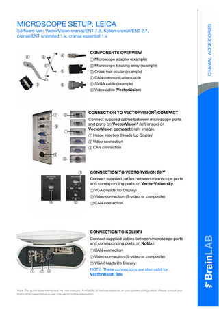
MICROSCOPE SETUP: LEICA
Software Ver.: VectorVision cranial/ENT 7.9, Kolibri cranial/ENT 2.7, cranial/ENT unlimited 1.x, cranial essential 1.x
a
COMPONENTS OVERVIEW
f
s
a Microscope adapter (example) s Microscope tracking array (example) d Cross-hair ocular (example)
g
f CAN communication cable d
g SVGA cable (example)
h
h Video cable (VectorVision)
a
CONNECTION TO VECTORVISION2/COMPACT
s
Connect supplied cables between microscope ports and ports on VectorVision² (left image) or VectorVision compact (right image).
s a
a Image injection (Heads Up Display) s Video connection d CAN connection
d d
a
CONNECTION TO VECTORVISION SKY Connect supplied cables between microscope ports and corresponding ports on VectorVision sky. a VGA (Heads Up Display) s Video connection (S-video or composite)
d
s
d CAN connection
CONNECTION TO KOLIBRI Connect supplied cables between microscope ports and corresponding ports on Kolibri. a CAN connection s Video connection (S-video or composite) d VGA (Heads Up Display) a
s d
NOTE: These connections are also valid for VectorVision flex.
Note: This guide does not replace the user manuals. Availability of features depends on your system configuration. Please consult your BrainLAB representative or user manual for further information.
DRAPING MICROSCOPE AND ADAPTER • Drape microscope as usual, including microscope adapter.
NOTE: Perform draping so that the surgeon can perform microscope navigation without compromising the sterile field.
ATTACHING STERILE REFERENCE ARRAY • Pierce drape and place sterile tracking array on adapter so that teeth of array match teeth of adapter. • Attach tracking array to adapter and tighten fixation screw. • Autobalance the microscope to compensate for weight changes due to the tracking array.
NOTE: You can attach the tracking array in the standard or 90° position, depending on the OR setup.
MANUFACTURER INFORMATION:
COPYRIGHT:
LIABILITY:
BrainLAB AG Kapellenstr. 12, 85622 Feldkirchen, Germany
This guide contains proprietary information protected by copyright. No part of this guide may be reproduced or translated without the express written permission of BrainLAB.
This guide is subject to change without notice and does not represent a commitment on the part of BrainLAB.
Europe, Africa, Asia, Australia: +49 89 99 15 68 44 USA & Canada: +1 800 597 5911 Japan: +81 3 3769 6900 Latin America: +55 11 3256-8301 France: +33-800-67-60-30 E-mail: [email protected]
Document Revision: 1.0 Article Number: 60903-68EN
For further information, please refer to the “Limitations of Liability” section in the BrainLAB Standard Terms and Conditions of Sale.
MICROSCOPE SETUP: MOELLER-WEDEL/OLYMPUS Software Ver.: VectorVision cranial/ENT 7.9, Kolibri cranial/ENT 2.7, cranial/ENT unlimited 1.x, cranial essential 1.x
COMPONENTS OVERVIEW a
s
f
a Microscope adapter (example) s Microscope tracking array (example) d Cross-hair ocular (example)
g
f Serial communication cable (example) g SVGA cable (example)
h
d
h Video cable (VectorVision)
CONNECTION TO VECTORVISION2/COMPACT
a s
Connect supplied cables between microscope ports and ports on VectorVision² (left image) or VectorVision compact (right image).
s a d
a Image injection (Heads Up Display) s Video connection
d
d Serial connection
s
a
CONNECTION TO VECTORVISION SKY Connect supplied cables between microscope ports and corresponding ports on VectorVision sky. a Serial connection s VGA (Heads Up Display)
d
d Video connection (S-video or composite)
CONNECTION TO KOLIBRI Connect supplied cables between microscope ports and corresponding ports on Kolibri. a Video connection (S-video or composite) s VGA (Heads Up Display) d Serial connection (back of Kolibri)
a s d
NOTE: These connections are also valid for VectorVision flex.
Note: This guide does not replace the user manuals. Availability of features depends on your system configuration. Please consult your BrainLAB representative or user manual for further information.
DRAPING MICROSCOPE AND ADAPTER • Drape microscope as usual, including microscope adapter.
NOTE: Perform draping so that the surgeon can perform microscope navigation without compromising the sterile field.
ATTACHING STERILE REFERENCE ARRAY • Pierce drape and place sterile tracking array on adapter so that teeth of array match teeth of adapter. • Attach tracking array to adapter and tighten fixation screw. • Autobalance the microscope to compensate for weight changes due to the tracking array.
NOTE: You can attach the tracking array in the standard or 90° position, depending on the OR setup.
MANUFACTURER INFORMATION:
COPYRIGHT:
LIABILITY:
BrainLAB AG Kapellenstr. 12, 85622 Feldkirchen, Germany
This guide contains proprietary information protected by copyright. No part of this guide may be reproduced or translated without the express written permission of BrainLAB.
This guide is subject to change without notice and does not represent a commitment on the part of BrainLAB.
Europe, Africa, Asia, Australia: +49 89 99 15 68 44 USA & Canada: +1 800 597 5911 Japan: +81 3 3769 6900 Latin America: +55 11 3256-8301 France: +33-800-67-60-30 E-mail: [email protected]
Document Revision: 1.0 Article Number: 60903-68EN
For further information, please refer to the “Limitations of Liability” section in the BrainLAB Standard Terms and Conditions of Sale.
MICROSCOPE SETUP: ZEISS Software Ver.: VectorVision cranial/ENT 7.9, Kolibri cranial/ENT 2.7, cranial/ENT unlimited 1.x, cranial essential 1.x
COMPONENTS OVERVIEW a
s
f
a Microscope adapter (example) s Microscope tracking array (example) d Cross-hair ocular (example)
g
f Serial communication cable (example) d
g SVGA cable (example)
h
h Video cable (VectorVision)
a
CONNECTION TO VECTORVISION2/COMPACT
s
Connect supplied cables between microscope ports and ports on VectorVision² (left image) or VectorVision compact (right image).
s a
d
a Image injection (Heads Up Display) s Video connection
f
f
d Handle connection (PC NV on Zeiss NC4)
d
f Serial connection
a
s
CONNECTION TO VECTORVISION SKY Connect supplied cables between microscope ports and corresponding ports on VectorVision sky. a Serial connection s VGA (Heads Up Display)
d
f
d Video connection (S-video or composite) f Handle connection (PC NV on Zeiss NC4)
CONNECTION TO KOLIBRI Connect supplied cables between microscope ports and corresponding ports on Kolibri. a Video connection (S-video or composite) s VGA (Heads Up Display) a s
d
d Handle connection (PC NV on Zeiss NC4) f Serial connection
f
NOTE: These connections are also valid for VectorVision flex.
Note: This guide does not replace the user manuals. Availability of features depends on your system configuration. Please consult your BrainLAB representative or user manual for further information.
DRAPING MICROSCOPE AND ADAPTER • Drape microscope as usual, including microscope adapter.
NOTE: Perform draping so that the surgeon can perform microscope navigation without compromising the sterile field.
ATTACHING STERILE REFERENCE ARRAY • Pierce drape and place sterile tracking array on adapter so that teeth of array match teeth of adapter. • Attach tracking array to adapter and tighten fixation screw. • Autobalance the microscope to compensate for weight changes due to the tracking array.
NOTE: You can attach the tracking array in the standard or 90° position, depending on the OR setup.
MANUFACTURER INFORMATION:
COPYRIGHT:
LIABILITY:
BrainLAB AG Kapellenstr. 12, 85622 Feldkirchen, Germany
This guide contains proprietary information protected by copyright. No part of this guide may be reproduced or translated without the express written permission of BrainLAB.
This guide is subject to change without notice and does not represent a commitment on the part of BrainLAB.
Europe, Africa, Asia, Australia: +49 89 99 15 68 44 USA & Canada: +1 800 597 5911 Japan: +81 3 3769 6900 Latin America: +55 11 3256-8301 France: +33-800-67-60-30 E-mail: [email protected]
Document Revision: 1.0 Article Number: 60903-68EN
For further information, please refer to the “Limitations of Liability” section in the BrainLAB Standard Terms and Conditions of Sale.
MICROSCOPE VERIFICATION & CALIBRATION Software Ver.: VectorVision cranial/ENT 7.9, Kolibri cranial/ENT 2.7, cranial/ENT unlimited 1.x, cranial essential 1.x
MICROSCOPE INITIALIZATION • Press the Tools button and select Microscope. • Press Connect Microscope. • In Tools > Microscope > Model (shown if more than one microscope available), select the microscope. • In the Microscope Detection dialog, select the correct setup depending on the tracking array position (standard or 90°).
VERIFICATION (REFERENCE ARRAY) Use this option to verify the microscope calibration using the reference array cone (divot). • Position microscope directly over reference array and focus microscope at maximum zoom on bottom of cone. • Verify that deviation (distance from navigated focal point to cone) is within an acceptable range. • Press Accept if accuracy is sufficient, or press Recalibrate to recalibrate microscope.
VERIFICATION (LANDMARKS) Use this option to verify the microscope calibration using anatomical or pre-planned landmarks. • Focus the microscope at maximum zoom on landmark. Press Video Verification to toggle to axial, coronal and sagittal views (optional). • Verify that deviation (distance from navigated focal point to landmark) is within an acceptable range. • Press Accept if accuracy is sufficient, or press Recalibrate to recalibrate microscope.
RECALIBRATION: POSITION MICROSCOPE • If recalibration is necessary, install the cross-hair ocular (if supplied). • Position microscope directly over reference array cone (divot).
NOTE: Position microscope as perpendicular as possible to plane of reference array.
Note: This guide does not replace the user manuals.
MICROSCOPE RECALIBRATION • Press Recalibration in the verification dialog, or press Calibrate Microscope in Tools > Microscope. • Focus microscope on reference array cone. • Ensure that focus is in optimal region (indicated by green area in dialog), and that cross-hair is focused and centered on bottom of cone. If necessary, finetune focus using focus buttons. • Press Recalibrate and verify the calibration.
MANUFACTURER INFORMATION:
COPYRIGHT:
LIABILITY:
BrainLAB AG Kapellenstr. 12, 85622 Feldkirchen, Germany
This guide contains proprietary information protected by copyright. No part of this guide may be reproduced or translated without the express written permission of BrainLAB.
This guide is subject to change without notice and does not represent a commitment on the part of BrainLAB.
Europe, Africa, Asia, Australia: +49 89 99 15 68 44 USA & Canada: +1 800 597 5911 Japan: +81 3 3769 6900 Latin America: +55 11 3256-8301 France: +33-800-67-60-30 E-mail: [email protected]
Document Revision: 1.0 Article Number: 60903-68EN
For further information, please refer to the “Limitations of Liability” section in the BrainLAB Standard Terms and Conditions of Sale.
MICROSCOPE FEATURES Software Ver.: VectorVision cranial/ENT 7.9, Kolibri cranial/ENT 2.7, cranial/ENT unlimited 1.x, cranial essential 1.x
MICROSCOPE NAVIGATION Following microscope verification, the Tools > Microscope > Options dialog opens. Close this dialog to begin microscope navigation. NOTE: See the Microscope Views page for information on viewing options.
IMAGE INJECTION Activate this option to superimpose contours of outlined objects displayed in navigation view into microscope’s field of view. • Press Options in Tools > Microscope, and select Image Injection. • In Tools > Microscope > Options, activate Color Overlay so that color of injected objects is defined by navigation system.
CLOSED SHUTTER Activate this option to close ocular view and display video view from navigation screen in microscope ocular. • In Tools > Microscope, press Options and select Closed Shutter.
SMART AUTO-FOCUS Activate this option to focus microscope exactly on tracked instrument tip (indicated by a cross-hair in the HUD). • In Tools > Microscope, press Options and select Smart Auto-Focus.
NOTE: The instrument tip must be within the microscope’s field of view.
Note: This guide does not replace the user manuals. Availability of features depends on your system configuration. Please consult your BrainLAB representative or user manual for further information.
SMART AUTO-TRACKING Activate this option to continuously track instrument position with microscope, focusing on instrument tip (indicated by a cross-hair in the HUD). The microscope stops tracking when it reaches the instrument tip. • In Tools > Microscope, press Options and select Smart Auto-Tracking.
FOCAL POINT/TARGET RETURN In Tools > Microscope, activate: • Go To Focal Point to return microscope to position and focus of prior stored focal point. • Store Focal Point to save the position and focal plane information of actual point. • Continuous Target Return to recall position and focal plane of most recently focused point (MoellerWedel only).
HANDLE CONTROL A C
D B
• Open Tools > Microscope and press Handle Control to access navigation functions available using microscope handle bars. • Use the handle bar controls as follows (example): - A: Previous (one menu item back) - B: Next (one menu item forward) - C: Cancel - D: OK (confirm an action). For Zeiss NC4 microscopes, D is in the center of the control.
HANDLE CONTROL - MENU STRUCTURE IN SOFTWARE Once you press Handle Control, the navigation menu is displayed in the microscope. • Use the handle control to scroll through the menu and activate the navigation features.
MANUFACTURER INFORMATION:
COPYRIGHT:
LIABILITY:
BrainLAB AG Kapellenstr. 12, 85622 Feldkirchen, Germany
This guide contains proprietary information protected by copyright. No part of this guide may be reproduced or translated without the express written permission of BrainLAB.
This guide is subject to change without notice and does not represent a commitment on the part of BrainLAB.
Europe, Africa, Asia, Australia: +49 89 99 15 68 44 USA & Canada: +1 800 597 5911 Japan: +81 3 3769 6900 Latin America: +55 11 3256-8301 France: +33-800-67-60-30 E-mail: [email protected]
Document Revision: 1.0 Article Number: 60903-68EN
For further information, please refer to the “Limitations of Liability” section in the BrainLAB Standard Terms and Conditions of Sale.
MICROSCOPE VIEWS Software Ver.: VectorVision cranial/ENT 7.9, Kolibri cranial/ENT 2.7, cranial/ENT unlimited 1.x, cranial essential 1.x
NAVIGATING MICROSCOPE AS POINTER Use default microscope navigation views to navigate the microscope as a virtual pointer.
a s d
• Blue line = microscope trajectory a. • Focus point of microscope = pointer tip s. • Blue ring = microscope’s field of view d (adjusts according to zoom factor).
MICROSCOPE DEPTHVIEW Open this view (eye icon > Other Views) to show anatomical patient data reconstructed to the corresponding live view in microscope oculars.
a
s d f
• Blue ring = microscope’s field of view a. • Blue cross-hair = center of focal plane s. • Solid contour line = border of planned object on this plane d. • Dotted contour line = extension of object above or below that plane f.
CONFIGURE MICROSCOPE VIDEO VIEW • Open Tools > Microscope and press Video Configuration. • In Input tab, press Input 1 or Input 2 (depending on the user panel to which microscope video is connected). • In Settings tab, adjust image view settings as needed. • Press Accept to confirm settings.
MICROSCOPE VIDEO VIEW
a s
Open this view (eye icon > Other Views > Microscope) to show contours of planned objects, trajectories, and labeled points overlaid onto a microscope video view. • Dotted contour line = extension of object above or below that plane a. • Solid contour line = border of planned object on this plane s.
Note: This guide does not replace the user manuals.
MANUFACTURER INFORMATION:
COPYRIGHT:
LIABILITY:
BrainLAB AG Kapellenstr. 12, 85622 Feldkirchen, Germany
This guide contains proprietary information protected by copyright. No part of this guide may be reproduced or translated without the express written permission of BrainLAB.
This guide is subject to change without notice and does not represent a commitment on the part of BrainLAB.
Europe, Africa, Asia, Australia: +49 89 99 15 68 44 USA & Canada: +1 800 597 5911 Japan: +81 3 3769 6900 Latin America: +55 11 3256-8301 France: +33-800-67-60-30 E-mail: [email protected]
Document Revision: 1.0 Article Number: 60903-68EN
For further information, please refer to the “Limitations of Liability” section in the BrainLAB Standard Terms and Conditions of Sale.