Carl Zeiss
OPMI pico Instructions for Use Issue 10.1 Oct 2005
Instructions for Use
58 Pages

Preview
Page 1
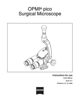
OPMI® pico Surgical Microscope
Instructions for use G-30-1384-en Issue 10.1 Printed on 27. 10. 2005
Key to symbols Different symbols used in this user's manual draw your attention to safety aspects and useful tips. The symbols are explained in the following.
Warning! The warning triangle indicates potential sources of danger which may constitute a risk of injury for the user or a health hazard.
Caution: The square indicates situations which may lead to malfunction, defects, collision or damage of the instrument.
Note: The hand indicates hints on the use of the instrument or other tips for the user.
OPMI®
OPMI® is a registered trademark of Carl Zeiss.
Contents –
Key to symbols
2
Functions at a glance
5
–
6
OPMI pico Surgical Microscope
Safety
7
–
Directives and standards
8
–
Notes on installation and use
9
Description
13
OPMI pico surgical microscope
14
–
Intended use
14
–
Controls and connections
16
OPMI pico with MORA interface.
18
–
Description
18
–
MORA interface with lateral output port for documentation
20
–
Attaching documentation accessories
20
OPMI pico with Endoport
22
Preparations for use
25
Attaching the equipment
26
–
Mounting the tubes, eyepieces and the objective lens
26
–
Mounting the handgrips
28
Connections
30
–
Mounting the light guide
30
–
Connecting the video camera
31
–
Connecting an endoscope camera or external digital camera
32
Operation
35
–
Adjusting the surgical microscope
36
–
Checklist
38
Procedure
G-30-1384-en
OPMI® pico Surgical Microscope
40
Issue 10.1 Printed on 27. 10. 2005
Maintenance / Further information
41
–
Trouble-shooting
42
–
Care of the unit
44
–
Sterilization
45
–
Disinfecting the control keys
46
–
Ordering data
47
–
Accessories
49
–
Technical data
50
–
Ambient requirements
51
Index
G-30-1384-en
OPMI® pico Surgical Microscope
53
Issue 10.1 Printed on 27. 10. 2005
5
Functions at a glance
Functions at a glance
OPMI pico Surgical Microscope
G-30-1384-en
OPMI® pico Surgical Microscope
6
Issue 10.1 Printed on 27. 10. 2005
6
Functions at a glance
OPMI pico Surgical Microscope 1 2 3 4 5 6 7
Connecting the light guide Selecting a filter Adjusting the interpupillary distance (push or pull) Setting your prescription Adjusting the eyecups Manual magnification adjustment Removing the cover and mounting the handgrip 8 Optional mounting of an accessory unit or handgrips 9 Connecting the system cable for the video camera 10 Locking / releasing the microscope's tilt motion
1 2
3
G-30-1384-en
4
5
6
7
8
OPMI® pico Surgical Microscope
9
page 30 page 16 ---page 16 page 28 page 28 page 31 page 16
10
Issue 10.1 Printed on 27. 10. 2005
Safety
7
Directives and standards
8
Notes on installation and use
9
Safety
G-30-1384-en
OPMI® pico Surgical Microscope
Issue 10.1 Printed on 27. 10. 2005
8
Safety
The instrument described in this manual has been developed and tested in accordance with Carl Zeiss safety standards and with national and international regulations. A high degree of instrument safety is thus ensured. We would like to inform you on the safety aspects involved in operating the instrument. This chapter contains a summary of the most important precautions to be observed. Further safety notes are also contained in other parts of this user's manual; they are marked with a warning triangle containing an exclamation mark as shown here. Please pay special attention to these safety notes. Safety is only ensured when this instrument is operated properly. Please read through this manual carefully before turning the instrument on. Also read through the user's manuals of the other equipment used with this instrument. You may obtain further information from our service organization or authorized representatives.
Directives and standards The system described in this user manual has been designed in compliance with: –
EN
–
UL
–
IEC
–
CSA
In accordance with Directive 93/42/EEC, the complete quality management system of the company Carl Zeiss, Surgery Products Division, has been certified by the DQS Deutsche Gesellschaft zur Zertifizierung von Managementsystemen mbH, a notified body, under registration number 250881 MP21.
G-30-1384-en
•
The instrument must be connected to a special emergency backup line supply in accordance with the regulations or directives which apply in your country.
–
As per Directive 93/42/EEC, the unit is a Class I instrument.
–
For USA: FDA classification Class I.
•
Please observe all applicable accident prevention regulations.
OPMI® pico Surgical Microscope
Issue 10.1 Printed on 27. 10. 2005
9
Safety
Notes on installation and use Safe working order •
G-30-1384-en
Do not operate the equipment contained in the delivery package in –
explosion-risk areas,
–
the presence of inflammable anesthetics or volatile solvents such as alcohol, benzine or similar chemicals.
•
Do not station or use the instrument in damp rooms. Do not expose the instrument to water splashes, dripping water or sprayed water.
•
Immediately unplug any equipment that gives off smoke, sparks or strange noises. Do not use the instrument until our service representative has repaired it.
•
Do not place any fluid-filled containers on top of the instrument. Make sure that no fluids can seep into the instrument.
•
Do not force cable connections. If the male and female parts do not readily connect, make sure that they are appropriate for one another. If any of the connectors are damaged, have our service representative repair them.
•
Potential equalization: The instrument can be incorporated into potential equalization measures. For this purpose, contact our service department.
•
Do not use a mobile phone in the vicinity of the equipment because the radio interference can cause the equipment to malfunction. The effects of radio interference on medical equipment depend on a number of various factors and are therefore entirely unforeseeable.
•
Modifications and repairs on these instruments or instruments used with them may only be performed by our service representative or by other authorized persons.
•
The manufacturer will not accept any liability for damage caused by unauthorized persons tampering with the instrument; this will also forfeit any rights to claim under warranty.
•
Use this instrument only for the applications described.
•
Only use the instrument with the accessories supplied. Should you wish to use other accessory equipment, make sure that Carl Zeiss or the equipment manufacturer has certified that its use will not impair the safety of instrument.
OPMI® pico Surgical Microscope
Issue 10.1 Printed on 27. 10. 2005
10
Safety
•
Only personnel who have undergone training and instruction are allowed to use this instrument. It is the responsibility of the customer or institution operating the equipment to train and instruct all staff using the equipment.
•
Keep the user's manuals where they are easily accessible at all times for the persons operating the instrument.
•
Never look at the sun through the binocular tube, the objective lens or an eyepiece.
•
Do not pull at the light guide cable, at the power cord or at other cable connections.
•
This instrument is a high-grade technological product. To ensure optimum performance and safe working order of the instrument, its safety must be checked once every 12 months. We recommend having this check performed by our service representative as part of regular maintenance work. If a failure occurs which you cannot correct using the trouble-shooting table, attach a sign to the instrument stating it is out of order and contact our service representative.
Requirements for operation Our service representative or a specialist authorized by us will install the instrument. Please make sure that the following requirements for operation remain fulfilled in the future: –
All mechanical connections (details in the user's manual) which are relevant to safety are properly connected and screw connections tightened.
–
All cables and plugs are in good working condition.
–
The voltage setting on the instrument conforms to the rated voltage of the line supply on site.
–
The instrument is plugged into a power outlet which has a properly connected protective ground contact.
–
The power cord being used is the one designed for use with this instrument.
Before every use and after re-equipping the instrument
G-30-1384-en
•
Make sure that all ”Requirements for operation” are fulfilled.
•
Go through the checklist.
•
Re-attach or close any covers, panels or caps which have been removed or opened.
OPMI® pico Surgical Microscope
Issue 10.1 Printed on 27. 10. 2005
11
Safety
•
Pay special attention to warning symbols on the instrument (triangular warning signs with exclamation marks), labels and any parts such as screws or surfaces painted red.
For every use of the instrument •
Avoid looking directly into the light source, e.g. into the microscope objective lens or a light guide.
•
Any kind of radiation has a detrimental effect on biological tissue.This also applies to the light illuminating the surgical field. Please therefore reduce the brightness and duration of illumination on the surgical field to the absolute minimum required.
Warning! The OPMI pico surgical microscope must not be used in ophthalmic examinations and surgery.
G-30-1384-en
OPMI® pico Surgical Microscope
Issue 10.1 Printed on 27. 10. 2005
12
Safety
G-30-1384-en
OPMI® pico Surgical Microscope
Issue 10.1 Printed on 27. 10. 2005
Description
13
OPMI pico surgical microscope
14
Intended use
14
Controls and connections
16
OPMI pico with MORA interface.
18
Description
18
MORA interface with lateral output port for documentation
20
Attaching documentation accessories
20
OPMI pico with Endoport
22
Description
G-30-1384-en
OPMI® pico Surgical Microscope
Issue 10.1 Printed on 27. 10. 2005
14
Description
OPMI pico surgical microscope Intended use The OPMI pico surgical microscope is a microscope for manual operation. It is a versatile instrument for daily routine work on seated and recumbent patients. You can use it as a –
surgical microscope
–
diagnostic microscope
–
training and dissecting microscope.
The OPMI pico surgical microscope can be combined with a wide range of accessories. The OPMI pico microscope is available –
without an integrated video camera
–
with an integrated video camera
–
with an integrated NTSC video camera
–
with an Endoport interface for endoscopes.
The endoscope port is used to attach an endoscope camera to OPMI pico. This allows you to transmit the image provided by OPMI pico to the video equipment of your endoscope system and display this image on its monitor and record it using a video recorder. Warning! The OPMI pico surgical microscope must not be used in ophthalmic examinations and surgery. Note: The OPMI pico i surgical microscope is available for the special needs of ophthalmology. OPMI pico and OPMI pico i have a different appearance: –
their lettering: OPMI pico or OPMI pico i
–
their color of the light guide socket: – OPMI pico: black light guide socket – OPMI pico i: light guide socket made of shiny aluminum
G-30-1384-en
OPMI® pico Surgical Microscope
Issue 10.1 Printed on 27. 10. 2005
15
Description
Note: This user's manual describes the instrument together with other units and accessories. The system described herein represents a common instrument configuration. The description generally applies for other similar configurations as well. The scope of the delivery package is not defined by the configuration shown in this manual, but by the delivery specification.
G-30-1384-en
OPMI® pico Surgical Microscope
Issue 10.1 Printed on 27. 10. 2005
16
Description
Controls and connections 1
Friction adjustment knob For locking the surgical microscope on the support arm.
2
Dust cover Remove this cover before mounting the binocular tube or another module on the surgical microscope.
3
Securing screw Screw in the securing screw as far as it will go after you have inserted the binocular tube or another module in the mount on the surgical microscope.
4
Magnification changer Use this knob to manually set the magnification to one of five clickstop positions (γ = 0.4x, γ = 0.6x, γ = 1.0x, γ = 1.6x, γ = 2.5x).
5
Cover You can remove this cover and mount a handgrip for microscope guidance instead.
6
Objective lens Objective lenses with different focal lengths are available, see ordering data. The focal length approximately corresponds to the working distance.
7
Holes for mounting accessories The bottom of the surgical microscope contains two holes mounting accessories (e.g. handgrip, dovetail mount).
for
8
Video connection socket for the system cable to the video camera (if integrated, option).
9
Light guide socket This socket is used to direct the light from the illumination system to the surgical microscope.
10 Filter selector Turn this selector to swing filters into the illumination beam path. There are three positions: no filter / green filter / orange filter – The green filter enhances contrast. – The orange filter prevents the premature hardening of composite fillings. 11 Support arm Supports the surgical microscope and is used for mounting it on a suspension system.
G-30-1384-en
OPMI® pico Surgical Microscope
Issue 10.1 Printed on 27. 10. 2005
17
Description
11 10
9 1
8
2 4
3 4 1
5 4 6 7
G-30-1384-en
OPMI® pico Surgical Microscope
Issue 10.1 Printed on 27. 10. 2005
18
Description
OPMI pico with MORA interface. The MORA interface is intended for use on the OPMI pico surgical microscope in dentistry and on the technoscope.
Description The MORA interface increases the microscope's mobility about its lateral tilt axis, without changing the user's upright, ergonomic seated posture. This offers advantages for the treatment of relatively large fields of therapy, e.g. when several teeth are being treated at once. A manually operated annular diaphragm permits the depth of field to be varied. Our service staff or an expert authorized by us will install the MORA interface for you. Please contact our service department or an authorized representative. Note: With maximum lateral tilt of the microscope body and high magnification, slight vignetting of the field of view may occur.
G-30-1384-en
1
Marking of zero position
2
Friction adjustment of lateral tilt movement The microscope body can be tilted by approx. 25° to the left or right, and can be locked in any position using the friction screw.
3
Locking screw Tighten this screw firmly after you have inserted the MORA interface in the mount on the OPMI pico microscope body.
4
Locking screw Tighten this screw firmly after you have inserted the binocular tube in the mount of the MORA interface.
5
Diaphragm setting knob A swing-in annular diaphragm permits you to increase the depth of field so that the field of therapy is in focus over a larger range. This involves a reduction in light intensity. Set the annular diaphragm to meet your specific requirements.
OPMI® pico Surgical Microscope
Issue 10.1 Printed on 27. 10. 2005
19
Description
Warning! Before every use and after re-equipping the instrument, make sure that the microscope body and binocular tube are securely locked in position. Make sure that locking screws (3) and (4) have been firmly tightened! Caution: • Please observe the maximum permissible load on the suspension system. With the S100 suspension systems, for example, this is 2.5 to 7.0 kg (complete microscope equipment including accessories and coupling)
1
2
3 4 5
ca ± 25°
G-30-1384-en
OPMI® pico Surgical Microscope
Issue 10.1 Printed on 27. 10. 2005
20
Description
MORA interface with lateral output port for documentation The MORA interface is optionally available with documentation equipment. This version is shown in the illustration on the opposite page. The camera adapter shown in the illustration is an example of further accessories that can be mounted on the documentation port. The method described below can also be used for other accessories. The operating principle of the accessories is described in the relevant user manuals.
Attaching documentation accessories •
Loosen knurled ring (2). Note: An arrow with the labeling "open" is located on the shaft next to knurled ring (2).
•
Remove dust cover (3) and store it in a safe place.
•
Insert camera adapter (4) in the opening of the image exit port. The opening of the image exit port is fitted with guide projections. Camera adapter (4) has the corresponding grooves. Carefully turn the accessory module until the guide projections fit into the grooves, and slide the camera adapter in the opening as far as it will go.
•
Screw knurled ring (2) onto accessory module (3).
•
Firmly tighten knurled ring (2).
Note: – If the camera adapter is mounted with the port pointing vertically upward, the image of the connected camera is displayed with a 90° rotation. To avoid this, you can mount the camera adapter rotated by 90° so that the port (incl. camera) points horizontally to the back. –
G-30-1384-en
Note the effect of annular diaphragm (1) swung into the beam path. We recommend that you swing the annular diaphragm out of the beam path for image capture to ensure that sufficient light is available. With the annular diaphragm swung into the beam path, the exposure time increases accordingly.
OPMI® pico Surgical Microscope
Issue 10.1 Printed on 27. 10. 2005