Guide Sheet
1 Page
Preview
Page 1
LEVITAN FPS SCOPE
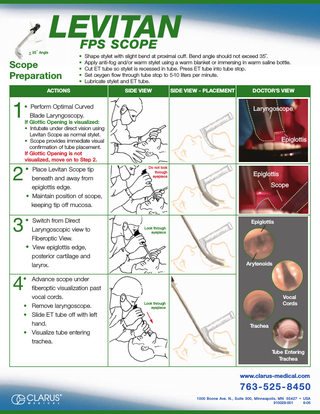
< 35˚ Angle
Scope Preparation
• Shape stylet with slight bend at proximal cuff. Bend angle should not exceed 35˚. • Apply anti-fog and/or warm stylet using a warm blanket or immersing in warm saline bottle. • Cut ET tube so stylet is recessed in tube. Press ET tube into tube stop. • Set oxygen flow through tube stop to 5-10 liters per minute. • Lubricate stylet and ET tube.
ACTIONS
1
SIDE VIEW
• Perform Optimal Curved Blade Laryngoscopy.
SIDE VIEW - PLACEMENT
DOCTOR’S VIEW
Laryngoscope
If Glottic Opening is visualized: • Intubate under direct vision using Levitan Scope as normal stylet. • Scope provides immediate visual confirmation of tube placement. If Glottic Opening is not visualized, move on to Step 2.
2
• Place Levitan Scope tip
3
• Switch from Direct
beneath and away from epiglottis edge. • Maintain position of scope, keeping tip off mucosa.
Laryngoscopic view to Fiberoptic View. • View epiglottis edge, posterior cartilage and larynx.
Epiglottis
Do not look through eyepiece
Epiglottis Scope
Epiglottis Look through eyepiece
Arytenoids
• Advance scope under
4
fiberoptic visualization past vocal cords. • Remove laryngoscope. • Slide ET tube off with left hand. • Visualize tube entering trachea.
Vocal Cords
Look through eyepiece
Trachea
Tube Entering Trachea
www.clarus-medical.com
763 - 525 - 8450 CLARUS M
E
D
I
C
A
L
®
1000 Boone Ave. N., Suite 300, Minneapolis, MN 55427 • USA 910029-001 6-05