IN TECH MEDICAL
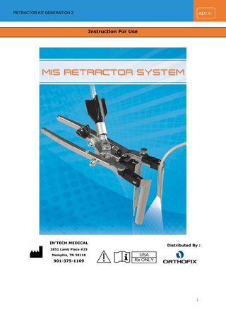
MIS retractor System
44 Pages

Preview
Page 1
REV A
RETRACTOR KIT GENERATION 2
Instruction For Use
IN’TECH MEDICAL 2851 Lamb Place #15
Distributed By :
Memphis, TN 38118
901-375-1109
1
REV A
RETRACTOR KIT GENERATION 2
Summary 1.
User manual ... 4 1.1.
1.1.1.
Destination ... 4
1.1.2.
Intended use ... 4
1.1.3.
Warnings ... 5
1.1.4.
Precautions ... 5
1.2.
Convenience kit content ... 6
1.2.1.
Cue card ... 6
1.2.2.
List of references and US/EU classes ... 8
1.2.3.
Example of a sequence of actions ... 9
1.3.
2.
Presentation ... 4
Specific handling instructions ...15
1.3.1.
Incision indicator ... 15
1.3.2.
Knife handle ... 15
1.3.3.
Dilators and dilator holder... 16
1.3.4.
Probe ... 17
1.3.5.
K-wire ... 18
1.3.6.
Retractor body, blades and wrench ... 18
1.3.7.
Table clamp and base... 23
1.3.8.
Disposable light mats and reusable extension cords ... 25
1.3.9.
Shim, broach and shim/broach holder ... 27
1.3.10.
Optional 4th blade and its support ... 28
1.3.11.
Penfield elevator and hockey stick ... 29
1.3.12.
Contrast puck... 30
1.3.13.
Stacking container ... 31
Instructions for cleaning, sterilization and maintenance ...32 2.1.
Examination ...32
2.1.1.
Visual inspection ... 32
2.1.2.
Functional inspection ... 32
2.2.
Handling prior to cleaning ...33
2.2.1.
Disassembling of the retractor ... 33
2.2.2.
Shim/broach holder disassembling ... 35
2.2.3.
4th blade support special handling care ... 35
2.2.4.
Dilators holder special handling care ... 36
2
REV A
RETRACTOR KIT GENERATION 2
2.2.5.
Table clamp base special handling care ... 36
2.2.6.
Dilators special handling care ... 36
2.3.
3.
Cleaning – decontamination ...37
2.3.1.
Preparation for cleaning... 37
2.3.2.
Manual pre-cleaning ... 37
2.3.3.
Automatic cleaning ... 37
2.4.
Sterilization ...39
2.5.
Storage ...39
2.6.
Maintenance ...40
2.6.1.
Retractor ... 40
2.6.2.
Table clamp connector... 40
2.6.3.
4th blade support ... 41
2.6.4.
Shim holder ... 41
2.7.
Complaints ...42
2.8.
Contact ...42
Chart of medical device symbols used ...43
3
REV A
RETRACTOR KIT GENERATION 2
1. User manual 1.1.
Presentation
1.1.1. Destination This kit is composed of several instruments which will be used by a trained orthopedic surgeon or neuro-surgeon to create access to the spine.
This kit is used to provide a spinal access channel through the tissue. The retractor will move apart the flesh to allow the surgeon access to the disc or at the wound bottom.
1.1.2. Intended use The Retractor Kit is intended to provide the surgeon with minimally invasive surgical access to the spine by ensuring the placement/positioning of the port, down to the bony spinal elements. These ports provide access to the spinal site which can be visualized using a microscope or loupes, and through which surgical instruments can be manipulated. DO NOT IMPLANT THE INSTRUMENTS.
4
REV A
RETRACTOR KIT GENERATION 2
1.1.3. Warnings
WARNING: Read the following handling instructions before use.
Breakage, misuse or mishandling of instruments, such as on sharp edges, may cause injury to the patient or the operative personnel. Improper maintenance, handling or poor cleaning procedures could render the device unsuitable for its intended use or even dangerous to the patient or surgical staff and void any warranty.
1.1.4. Precautions -
-
Extreme care should be taken to ensure that this instrument remain in good working order. o
Small parts can be lost.
o
Some instruments are sterile (probe and light mat): check that the packaging is unharmed. Check the expiry date on the sterile products.
o
Some parts are sharp and require to be handled with care in order not to harm the patient and the medical staff.
o
Devices must be handled with care to prevent damage. Take precautions to prevent any breakage. Instruments should not be bent or damaged in any way.
The user of this product must be familiar and trained in use and care of the product.
-
Do not use this instrument for any action for which it was not intended.
-
Before the surgery, to avoid injury, always run an examination as described below. If anything is missing or doesn't seem right, call your local agent as soon as possible and discard the kit.
-
Devices, that are provided non-sterile, must be cleaned and sterilized prior to use according to the directions outlined below (except otherwise stated).
-
Be aware that any failure in cleaning, maintenance or usage can lead to an unusable, corroded, broken instrument that could be dangerous to the patient and the medical staff.
5
REV A
RETRACTOR KIT GENERATION 2
1.2.
Convenience kit content
1.2.1. Cue card Below are pictures of the loaded Retractor Kit. Content might be custom so the pictures below are for information only. Custom Cue Card will be included in the packaging.
Figure 1: Case 1 - Upper level
Figure 2: Case 1 – Lower level
6
REV A
RETRACTOR KIT GENERATION 2
Figure 3: Case 2 – Upper level
Figure 4: Case 2 – Lower level
7
REV A
RETRACTOR KIT GENERATION 2
Wrench OFIX-208 OFIX-319
Disposable Probe (Sterile) S06ITM231
Disposable long light mat (Sterile) S06ITM308
Dual Extension Cord for Light Mat S06ITM234 Retractor with asymmetric body OFIX-271 Knife handle (Knurled) OFIX-272 Contrast puck OFIX-276 Incision indicator OFIX-277 4th blade 12x160mm Ti OFIX-284 4th blade attachment 12-18mm OFIX-286 Rotative table clamp base S06ITM288
CE Class
Shim and Broach Inserter OFIX-204
510(k)
Shim OFIX-201
Nonsterile
FDA class
Broach OFIX-200
Singleuse / reusable
K-wire OFIX-189 Blade 100mm OFIX-195-100 Blade 110mm OFIX-195-110 Blade 120mm OFIX-195-120 Blade 130mm OFIX-195-130 Blade 140mm OFIX-195-140 Blade 150mm OFIX-195-150 Blade 160mm OFIX-195-160 Table clamp (arm) OFIX-197
Sterile / Nonsterile
1.2.2. List of references and US/EU classes
Reusable
I
NA
I
Nonsterile
Reusable
I
NA
IIa
Reusable
I
NA
I
Reusable
I
NA
IIa
Reusable
I
NA
IIa
Reusable
I
NA
I
Reusable
I
NA
I
Reusable
I
NA
I
II
K063729
III
I
NA
III
Reusable
II
K901035
I
Reusable
I
NA
I
Reusable
I
NA
I
Reusable
I
NA
I
Reusable
I
NA
I
Reusable
I
NA
IIa
Reusable
I
NA
I
Reusable
I
NA
I
Nonsterile Nonsterile Nonsterile Nonsterile Nonsterile Nonsterile
Single-use only Sterile
Single-use only Sterile
Nonsterile Nonsterile Nonsterile Nonsterile Nonsterile Nonsterile Nonsterile Nonsterile
8
REV A
Sterile / Nonsterile
Singleuse / reusable
FDA class
510(k)
CE Class
RETRACTOR KIT GENERATION 2
Nonsterile
Reusable
I
NA
I
Reusable
I
NA
I
Reusable
I
NA
I
Dilator #1 OFIX-292-01 Dilator #2 OFIX-292-02 Stacking tray OFIX-295-2
Nonsterile Nonsterile
Penfield OFIX-273
1.2.3. Example of a sequence of actions This documentation does not provide medical advice. The content is not intended to be a substitute for professional training regarding the medical devices or procedure depicted herein. The following sequence of actions is for informational purpose only. Step 1: incision, dilators and nerve monitoring
Figure 5
Figure 6
Fig. 7: First spot where the incison should be by using the incision locator to locate the disc via fluroscopy. Then make an incision with a knife handle and a blade. Insert smallest dilator in the incision. Hold steady the dilator with the dilator holder during fluoroscopy.
9
REV A
RETRACTOR KIT GENERATION 2
Figure 7
Fig. 8 & 9: If desired for a particular approach to the spine, a nerve monitoring probe can slide down the small dilator to detect nerve(s) all around the small dilator. After nerve monitoring, remove the probe and insert the k-wire onto the disc.
Figure 8
Figure 9
10
REV A
RETRACTOR KIT GENERATION 2
Fig. 10 & 11: Slide the biggest dilator onto the small one. Run nerve monitoring again if desired for surgical access. Probe can slide down the big dilator dedicated channel to allow the probe to detect nerve all around the big dilator.
Figure 10
Figure 11
Fig. 12: Remove the probe. The scale on the tube will help chosing the required blade length.
Figure 12
11
REV A
RETRACTOR KIT GENERATION 2
Step 2: distraction and fastening system Fig 13 & 14 & 15: Take the retractor body and connect 3 blades: 1 right, 1 left, 1 central. They should make a general circle with small gaps between the blades. Slide the 3 blades circle down the dilator.
Figure 13
Figure 14
Figure 15
Fig 16 & 17: Fasten the retractor body to the surgery table with the articulated arm and its table clamp. Then remove the dilators and k-wire. Single-use light mat can be used to bring light to the bottom of the wound: slide the light mat down the blade. Probe can be used again for nerve monitoring if needed.
Figure 16
Figure 17
12
REV A
RETRACTOR KIT GENERATION 2
Fig. 18 & 19 & 20 & 21: To distract left and right blades, press both arms. Secure by screwing the lateral knob. To distract the central blade, turn the central black knob. This will allow to enlarge the access. Lateral blades can even be angled using the additional wrench.
Figure 21
Figure 18 Figure 19
Figure 20
Fig. 22 & 23: Remove the arms to save some space around the access.
Figure 22
Figure 23
13
REV A
RETRACTOR KIT GENERATION 2
Fig. 24 & 25: Fasten a broach onto the shim/broach inserter. Slide the broach down the blade via the dedicated groove. Insert the broach onto the disc, slight impacts on the back of the shim holder might help. Same procedure can be done to insert a shim onto the vertebral body. This will allow user to fasten the retractor body to the body.
Figure 24
Figure 25
Step 3: Additional tools Fig. 26 & 27 & 28: If a 4th blade is required, clip a 4th blade support on the top of the retractor. This support’s arms can slide to mates with the retractor opening. Slide a 4 th blade down the support’s dedicated slot. Lock its position with the additional wrench.
Figure 26
Figure 27
Figure 28
A contrast puck is also available. Refer to the individual product description to know more about these devices. All the reusable devices can be stored inside the dedicated container, which can also be sterilized. 14
REV A
RETRACTOR KIT GENERATION 2
1.3.
Specific handling instructions
1.3.1. Incision indicator Incision indicator is reusable. It can be used to locate the incision. Place the incision indicator cross above the incision. The device will be visible under fluoroscopy.
Figure 29
It must be handled with care to prevent damage. Take precautions to prevent tip breakage. 1.3.2. Knife handle The Knurled Knife handle is reusable.
Figure 30
Clip the scalpel blade onto the knife handle. Warning: scalpel blades are very sharp. They must be handled with care.
15
REV A
RETRACTOR KIT GENERATION 2
Figure 31
1.3.3. Dilators and dilator holder Dilators and dilator holder are reusable. Slide the smallest dilator in order to extend the incision. Slide carefully the largest dilator on top of the smallest dilator in order to extend even more the incision. Once the largest dilator is in position, it will provide the surgeon with the desired diameter where the retractor blades will fit tightly around it for the insertion of the device. Use the dilator holder to hold the dilators (see figure 32) while performing fluoro. Dilator holder can be fastened to the table clamp to avoid surgeon hand exposure during X-rays.
Figure 32
When fully inserted, the scale on the dilator will provide an approximate depth which is a hint for the selection of the blades length.
16
REV A
RETRACTOR KIT GENERATION 2
1.3.4. Probe The disposable probe is a sterile single use product. Check the expiry date and the packaging before use. Dispose after use. WARNING: Read the own Instruction For Use of the probe before any use of the product. In’Tech can't be responsible for any misuse of the disposable probe. This Instruction for Use is included inside the packaging.
Plug the probe to a suitable electrical source via DIN 42802 touch proof connectors. The probe can be carefully inserted in the dilator offset groove. The detection ball (located at the end of the probe) will be exposed at the end of the dilator (see next figure). The ball probe should never stick out from the distal end of the dilator probe. Do not overstrain nor press too hard while inserting the probe. When monitoring is done, carefully remove the probe from the dilator. Dispose the probe after use.
Figure 33
17
REV A
RETRACTOR KIT GENERATION 2
1.3.5. K-wire The K-wire is a reusable product. Be careful, K-wire is very sharp. Wearing gloves is recommended.
1.3.6. Retractor body, blades and wrench The retractor body, blades and wrench are reusable devices. The retractor body is available in an asymmetrical version were both arms can be open independently from each other. Blades are available with many lengths. - Choose the blades size based on the information provided by the scale on the dilators (blades length depending on the thickness of the patient) - Fix the 3 selected blades on the retractor body: they slide vertically either from the top or from beneath, until a click can be heard:
Place a right blade (R marked) on the right arm (R marked) (see next figure)
Place a left blade (L marked) on the left arm (L marked)
Place a central blade (C marked) on the middle arm
- Before inserting the retractor in the wound, check that the retractor is well closed. (see figure 34. Note: Image 34 is for illustration only and may vary from the actual product. There will be a small gap between the blades when in the closed position). If necessary, screw the right and left knobs using the wrench to set back to 0 the angle of the blades (refer to figure 38 for more details).Then slide down the retractor body with its blades in the wound around the largest dilator (see figure 35).
18
REV A
RETRACTOR KIT GENERATION 2
R
L L
R
C
Figure 34
Figure 35
- Spreading is obtained via 3 different movements (see figures below):
a) Spreading of the lateral blades: Squeezing the handle and screw the knob to maintain spreading as needed (see figure 37).
b) Tilting movement of the lateral blades: to enlarge the space on the bottom, use the screws on the left and right arms: screwing opens angularly the blades, unscrewing closes the blades (see figure 38).
c) Lateral movement of the selected blades: Screwing the big black knob will enlarge the opening, either the central blade will slide posterior or the 2 lateral blades will slide anterior, depending on where the retractor clamp is fixed to the table (see figure 39).
19
REV A
RETRACTOR KIT GENERATION 2
c) Knob for central blade: a) Knob for spreading the lateral blades
Lateral movement of the selected blades
b) Knob for tilting the lateral blades
Figure 36
Figure 37
Figure 38
Figure 39
20