Laerdal Medical
Laerdal Airway Management Trainer Directions for Use Rev C
Directions for Use
28 Pages

Preview
Page 1
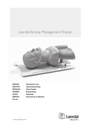
Laerdal Airway Management Trainer
ENGLISH DEUTSCH FRANÇAIS SVENSKA SUOMI ESPAÑOL Part list
Directions for Use Gebrauchsanweisung Mode d’emploi Bruksanvisning Käyttöohje Instrucciones de utilización
www.laerdal.com
facilitate tube insertion and prevent regurgitation of stomach contents. Laryngospasm can be simulated by instructor using a syringe with slide lock on the connection tubing.
The Laerdal Airway Management Trainer is mounted on a practice board and stored in a carrying case. The following equipment is provided:
Press on syringe piston to simulate laryngospasm. Maintain spasm using slide lock.
1. Sanitation kit: a) Container w/lid b) Compresses (10 ea) c) Syringe (60 ml) d) Cleaning pump assembly w/triple connector 2. Concentrated simulated vomit (150 grams) 3. Airway lubricant 4. Airway anatomy demonstration model
Vomiting can be simulated. See instructions for simulated stomach contents on bottle. See instructions under “Suction” for filling stomach and inducing unexpected vomiting. Excessive laryngoscope pressure on upper teeth is signalled by an audio signal. Proper tube placement can be checked by: - visual inspection of lung expansion during ventilation - auscultation of breathing sounds via built-in diaphragms
Applications The Laerdal Airway Management Trainer realistically simulates a non-anesthetized patient. It can be used to demonstrate and practice intubations, ventilation, suction and bronchoscopy. Intubation
- spontaneous breathing can be simulated by rhythmically squeezing and releasing lungs. The audible movement of air helps intubator locate tube position - bronchoscopic evaluation of tip position Complete training in all intubation procedures, including preoxygenation, can be practiced: - tracheal (oral and nasal) - pharyngeal (oral and nasal) - esophageal - bronchial – with optional bronchial tree
The airway anatomy demonstration model features realistic details and can be used to train recognition of landmarks. Suitable for demonstrating cricoid pressure.
Use lubricant on tube and in airway before inserting tube. Cricoid pressure can be used realistically to 2
ENGLISH
Ventilation
The Laerdal Airway Management Trainer allows use of all normal maneuvers to maintain an open airway; head tilt, chin lift, neck lift, jaw thrust.Ventilation can be practiced with or without equipment; mouth-tomouth, mouth-to-nose, mouth-to-mask, bag-valvemask.
Stomach contents can be effectively removed from the interior of the trainer by flushing with clean water immediately after training session. Even stomach contents that have unintentionally been allowed to dry inside the trainer, will redissolve in water. To reduce dissolution time circulate water through the head using the cleaning pump assembly. See “Sanitation after training “ point (B) below.
The trainer’s exterior and interior can be effectively cleaned after exhaled air resuscitation. (See under “Sanitation”)
Bronchoscopy Ventilation with excessive volume, or attempted against an obstructed airway, will cause inflation of the stomach. Make sure the elastic band is in place on the stomach retention valve. Note: Manikin exhales through mouth and nose.
A realistic bronchial tree is available as an option. It reproduces the bronchial airway down to the third generation. Both rigid and flexible fiberoptic bronchoscopy can be performed in order to learn - landmark recognition - proper instrument handling - bronchoscopic checking of tracheal and bronchial tube position - removal of solid material and mucus - identifying common pathologies
Suction The Laerdal Airway Management Trainer can be used for training in clearing the obstructed airway by suctioning liquid matter from: - oral cavity - oro- or nasopharynx - oro- or nasotrachea, via endotracheal tube - bronchial, using the optional bronchial tree Gastric drainage may also be practiced.
The bronchial tree is hidden from the student’s view during practice.The end of the bronchoscope transilluminates the airway so that the instructor can easily locate bronchoscope position during endoscopy. The bronchial tree consists of detachable left and right branches mounted on the bronchi of a separate trachea piece.This permits demonstration of the bronchial tree as a separate unit. For use on the trainer, detach both branches and mount them on the bronchi of the trainer, following color coded/ position marks.
Prepare simulated vomit. See mixing instructions on bottle. Remove the stomach retention valve. Fill stomach with approx. 4 cups of simulated vomit using a funnel inserted into the rigid connector.
Remount retention valve and connect stomach to esophagus.To induce vomiting press on the full stomach. 3
Sanitation
B. If exhaled air resuscitation has been practiced on the trainer, disinfection of the airways is required before storage or the next training session. Use the set-up described below and carry out the following four step procedure, changing liquid for each new step: 1. Soapy water to remove condensation on interior surfaces. 2. Clean water to remove soap residue. 3. Disinfecting solution. Allow to remain in completely filled airways for at least 10 min. 4. Clean water to remove disinfecting solution.
Open the practice board lock and lift the trainer off the board.
Place the trainer’s head face down, diagonally in the sanitation kit container filled with liquid to a point just over the internal ridge. Attach stomach and lung connectors to the triple connector of the cleaning pump assembly.
Disconnect lung tube connectors and remove lungs.
Detach and remove stomach.
Put the free tube end into the container. Place valve in one of the fastening brackets in the rear of the trainer’s shoulders. Insert the syringe into the opening of the valve.
A. When simulated vomit has been used, hold trainer over sink with tap water running into its mouth to flush residue from stomach and lung tubing. Shake and allow to drip dry. Circulate liquid through the airways by pumping the syringe plunger. After each step lift face clear of liquid to allow drainage.
Unscrew outer part from inner part of lung connecting piece. Flush bags. Disconnect stomach retention valve and open closure at other end of stomach. Flush stomach. Allow all parts to air dry before reassembly. 4
ENGLISH
Lungs and stomach Disassemble as described in (A) above. Pour liquids in the suggested order into stomach and each lung.
Shake well. Disinfecting solution should remain for at least 10 min.Wash couplings and submerge in disinfecting solution for at least 10 min. Flush in clean water. Allow all parts to dry before reassembly on trainer.
Preventive care Avoid contact between manikin’s skin and materials such as painted or lacquered surfaces, newsprint, ballpoint pens and lipstick. Advise trainees to remove lipstick and wash hands. To wash the manikin’s skin, use warm water and a mild detergent. Problem stains may be removed with alcohol if treated immediately. Excessive force has no place during endotracheal intubation. Strong force can cause severe trauma to the patient, and may also damage the plastic “tissue” of the manikin.
5
Parts
25 00 00
04 20 00 25 01 00 25 02 00 25 03 00 25 04 00 25 05 00 25 06 00 25 07 00 15 07 04 25 09 00 25 10 00 25 11 00 25 12 00
Airway Management Trainer, complete Parts Carrying case Foam block holder Head protector Practice board, cpl. w/lock Left lung Support plate for left lung Right lung Support plate for right lung Lung connecting piece (female) w/nut Left lung tube w/connectors Right lung tube w/connectors Stomach, complete Laryngospasm simulator,cpl
20 02 13 25 13 00 25 14 00 25 15 00 25 16 00 25 17 00 25 18 00 25 19 00 25 20 00 25 20 10 25 21 00 25 22 00 25 23 00
Y-piece (pkgs 10) Shoulder bracket Outer shoulder flange w/screws Inner shoulder flange w/7 screws and 2 rubber stoppers Shoulder piece Sound diaphragm and tubing Revolving shoulder disk Shoulder skin Head skin and airways cpl. w/teeth Teeth and audio signal device for tooth pressure Larynx cpl. w/screws and spasm device Skull, complete Lower jaw
26
25 24 00 25 25 00 25 26 00 25 28 00 25 20 90 25 30 00
25 90 00
Neck, complete Airway anatomy demonstration model Cleaning kit, cpl Concentrated simulated vomit Airway lubricant Manual for Airway Management Trainer Optional equipment Bronchial tree, complete
© 2005 Laerdal Medical AS, Alll Rights Reserved. 6511 rev C Printed in Norway