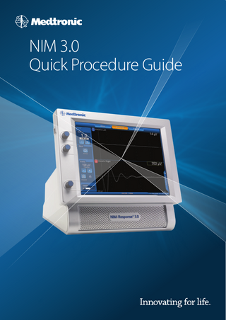
Quick Procedure Guide
40 Pages

Preview
Page 1
NIM 3.0 Quick Procedure Guide
This Quick Start Guide is only intended as a Quick Reference Manual of the NIM Response ®3.0. For more detailed information please consult the Product Manual. Created by the 2015 European & Canadian NIM specialist team
THIS ICON IS DEDICATED TO ANESTHESIOLOGISTS
NIM 3.0 QUICK PROCEDURE GUIDE
Plug & Check
Connections 1. Power cord 2. Interface 3. Muting clamp 4. Power switch ON ARE MANDATORY
PRINTER
2
PATIENT INTERFACE 8 OR 4 CHANNELS
USB KEY NEVER PLUG the USB key before turning on the NIM
4
POWER SWITCH ON/OFF
3
1
MUTING DETECTOR
POWER CORD
SURGEON MINI SCREEN
N I M 3 .0 Q U I C K P R O C E D U R E G U I D E
Plug & Check
System Quick Check with Simulator The check confirms that all electrodes circuits are connected and functioning properly. (For more details see Product Manual)
EX: 4 CHANNELS
NIM 3.0 QUICK PROCEDURE GUIDE
1.
Turn on the NIM
2.
In SET-UP, select a specific surgery
3.
Following the picture on the screen, connect all color coded cables (simulated subdermal electrodes, Ground and Stim 1 return) to the corresponding patient interface
4.
Connect a monopolar probe to the Stim 1 jack
5.
Automatic electrode check: full the connection
6.
Go to MONITORING menu. Touch and hold the probe to channel 1 of the stimulator and observe a typical EMG curve associated with an audible alarm. Repeat test for all channels involved in this specific surgery.
7.
You must obtain an EMG curve on the NIM screen and an audible alarm
8.
At the end of the complete process, turn off the NIM
3confirms integrity of
Facial Nerve 2 channels Main procedures: Otology Otospongiosis Mastoidectomy Tympanoplasty Cholesteatoma Cochlear implant
Otology
Setup Turn on the NIM After a system quick check, place the NIM 3.0 within the surgeon’s view and away from the electrocautery unit. Select Neuro/Otology on the screen. Choose the specific procedure from the drop down menu (Ex: Mastoid, Cochlear Implant).
Required disposables: Paired Subdermal Electrodes 2 channels REF 8227410 Other:………………………………………………………. Prass Standard Monopolar Probe Ref 8225101 Other:……………………………………………………….
Precaution for anesthesia: Do not use long-term paralyzing anesthetics to ensure proper EMG monitoring.
N I M 3 .0 Q U I C K P R O C E D U R E G U I D E
Prepare skin and place (subdermal) electrodes Insert green Ground electrode into the sternum, and then the white & red Stim Return (+) below. Keep a 2 cm distance between the two needles. Be sure to properly fix the electrodes or needles with an adhesive surgical tape. Connect all the wires to the patient interface box respecting color code.
Otology
Automatic Electrode Check Green check: Test passed. The integrity of the connection is validated by confirming the impedance values of the electrodes are within expected range. Red cross: Test failed. Replace electrode in question and check connection to patient interface box.
When everything is green, start your sterile field. Go to Monitoring tab.
Monitoring Clip Muting Detector to monopolar electrocautery cord(s). Do not include grounding pad or bipolar cord(s).
Connect the stimulating probe to the patient interface (STIM 1 black jack).
NIM 3.0 QUICK PROCEDURE GUIDE
Use the probe and keep it on the nerve for at least 1 sec. A typical EMG curve is • a white biphasic curve • preceded by a latency time • with a significant amplitude • accompanied with an audible alarm
Default settings Stimulation: 0,8 mA Event threshold: 100 microvolts On request of the surgeon, you can increase or decrease those levels
Optional Take a snapshot and capture the EMG curve by touching the quick tab at the bottom of the screen. For an academic reporting, follow this sequence: 1. FN Pre for Facial Nerve Pre Dissection 2. FN Post for Facial Nerve Post Dissection The snapshots are saved for the duration of the procedure. (It allows you to print or save a report on a USB stick. See next step.)
N I M 3 .0 Q U I C K P R O C E D U R E G U I D E
Otology
Stimulate the nerve with the probe pre and post dissection.
Reports Save or print your event report. Select information.
Otology
Enter patient information in the white box. A keyboard appears by touching the screen.
Validate with OK and press the arrow.
Select Quick Report and enter a USB Key.
Select All snapshots/ 4 per pages. Press Save. Do not disconnect during the saving process. Remove your USB key. Turn off the NIM.
NIM 3.0 QUICK PROCEDURE GUIDE
Facial Nerve 4 channels Main procedures Acoustic neuroma Parotidectomy
Setup Turn on the NIM After a system quick check, place the NIM 3.0 within the surgeon’s view and away from the electrocautery unit.
Parotid / Acoustic neuroma
For acoustic Neuroma: Select Neuro/Otology on the screen and choose Acoustic Neuroma (4 ch) from the drop down menu. For Parotid: Select Head/Neck on the screen and choose Parotid (4 ch) from the drop-down menu. Parotid in 4 channel as reference by “Clinical Outcome of Continuous Facial Nerve Monitoring During Primary Parotidectomy Jeffrey E. Terrell, MD; Paul R. Kileny, PhD; Chris Yian, MD; Ramon M. Esclamado, MD; Carol R. Bradford, MD; Matthew S. Pillsbury; Gregory T. Wolf, MD. Required disposables: Paired Subdermal Electrodes 4 channels REF 8227411 Other:………………………………………………………. Prass Standard Monopolar Probe Ref 8225101 Other:……………………………………………………….
Precaution for anesthesia: Do not use long-term paralyzing anesthetics to ensure proper EMG monitoring. N I M 3 .0 Q U I C K P R O C E D U R E G U I D E
Prepare skin and place (subdermal) electrodes. Insert green Ground electrode into the sternum, and then the white & red Stim Return (+) below. Keep a 2 cm distance between the two needles. Be sure to properly fix the electrodes or needles with an adhesive surgical tape. Connect all the wires to the patient interface box respecting color code.
Automatic Electrode Check. Green check: Test passed. The integrity of the connection is validated by confirming the impedance values of the electrodes are within expected range. Red cross: Test failed. Replace electrode in question and check connection to patient interface box.
Parotid / Acoustic neuroma
When everything is green, start your sterile field. Go to Monitoring tab.
Monitoring Clip Muting Detector to monopolar electrocautery cord(s). Do not include grounding pad or bipolar cord(s).
Connect the stimulating probe to the patient interface (STIM 1 black jack).
NIM 3.0 QUICK PROCEDURE GUIDE
Use the probe and keep it on the nerve for at least 1 sec. A typical EMG curve is • a white biphasic curve • preceded by a latency time • with a significant amplitude • accompanied with an audible alarm
Stimulate the nerve with the probe pre and post dissection.
Default settings Stimulation: 0,8 mA Event threshold: 100 microvolts
Parotid / Acoustic neuroma
On request of the surgeon, you can increase or decrease those levels
Optional Take a snapshot and capture the EMG curve by touching the quick tab at the bottom of the screen. For an academic reporting, follow this sequence: 1. FN Pre for Facial Nerve Pre Dissection 2. FN Post for Facial Nerve Post Dissection The snapshots are saved for the duration of the procedure. (It allows you to print or save a report on a USB stick. See next step.)
N I M 3 .0 Q U I C K P R O C E D U R E G U I D E
Reports Save or print your event report. Select information.
Enter patient information in the white box. A keyboard appears by touching the screen.
Validate with OK and press the arrow.
Select Quick Report and enter a USB Key.
Parotid / Acoustic neuroma
Select All snapshots/ 4 per pages. Press Save. Do not disconnect during the saving process. Remove your USB key. Turn off the NIM.
NIM 3.0 QUICK PROCEDURE GUIDE
Vagus & Recurrent Nerves (Superior Laryngeal Nerve) Main procedures Thyroidectomy Parathyroidectomy Neck dissection
Setup
Turn on the NIM After a system quick check, place the NIM 3.0 within the surgeon’s view and away from the electrocautery unit. Select Head/Neck on the screen and choose Thyroid from the drop-down menu .
Required disposables: Prass Standard Monopolar Probe Ref 8225101 Other:………………………………………………………. EMG Endotracheal Tube Ref 8229xxx
Thyroid
Other:……………………………………………………….
N I M 3 .0 Q U I C K P R O C E D U R E G U I D E
EMG Endotracheal Tubes The NIM EMG tube monitors electromyography activity during surgery and functions as a normal endotracheal tube, except that it contains bipolar electrodes for continuously monitoring both vocal cords during surgery. I.
Do not use long-term anesthetics to ensure proper EMG monitoring. II. In case of hesitation between two sizes, prefer the larger one for a better contact with vocal cords. III. Lubricate the cuff with a non paralysing aquous lubricant . IV. Follow specific instructions for each tube model (markers). V. Visual check for a perfect tube position (depth and rotation). Ideally with a standard or video laryngoscope. VI. Keep the tube close to midline and secure the tube with tape. VII. Check impedance after complete patient set-up.
PROPER POSITIONING TRIVANTAGE Blue cross at the vocal cords 4 Silver ink electrodes Black numbers and cuff anterior (12h), median Unique reference for Superior Laryngeal Nerve monitoring
FLEX TUBE Use of a stylet often required Symmetric electrodes (double black bands) against vocal cords – 2/3 behind VF/ 1/3 before Cuff anterior (12h), median
STANDARD REINFORCED
Thyroid
Blue numbers anterior (12h), median Electrodes (double metallic band in the blue rectangle) against vocal cords-2/3 behind VC/ 1/3 before
NOTE: The correct positioning of the tube is vital to achieve a good EMG response.
NIM 3.0 QUICK PROCEDURE GUIDE
Prepare skin and place (subdermal) electrodes. Insert green Ground electrode into the sternum, and then the white & red Stim Return (+) below. Keep a 2 cm distance between the two needles. Be sure to properly fix the electrodes or needles with an adhesive surgical tape. Connect all the wires to the patient interface box respecting color code.
Automatic Electrode Check. Green check: Test passed. The integrity of the connection is validated by confirming the impedance values of the electrodes are within expected range. Red cross: Test failed. Replace electrode in question or tube and check connection to patient interface box. Press Show details.
Impendance provides you additional information about electric loop/connectivity. Try to bring up the standard value. Values change instantly when you move the tube. An additional visual check is essential for a proper tube positioning (electrodes against vocal cords).
More info in the Troubleshooting guide
When everything is green, go to Monitoring tab.
Monitoring
Thyroid
Clip Muting Detector to monopolar electrocautery cord(s). Do not include grounding pad or bipolar cord(s).
N I M 3 .0 Q U I C K P R O C E D U R E G U I D E
Connect the stimulating probe to the patient interface (STIM 1 black jack).
Use the probe and keep it on the nerve for at least 1 sec.
A typical EMG curve is • a white biphasic curve • preceded by a latency time • with a significant amplitude • accompanied with an audible alarm
Stimulate the nerve with the probe pre and post dissection.
Default settings Stimulation: 1.0 mA Event threshold: 100 microvolts On request of the surgeon, you can increase or decrease those levels
Optional Take a snapshot and capture the EMG curve by touching the quick tab at the bottom of the screen. For an academic reporting, follow this sequence: (Ex for Left side) 1. 2. 3. 4.
Left
Right
L-V1 Vagus Nerve Pre Dissection Left L-R1 Recurrent Laryngeal Nerve Pre Dissection Left L-R2 Recurrent Laryngeal Nerve Post Dissection Left L-V2 Vagus nerve Post Dissection Left
L-S1 Superior Laryngeal Nerve Pre Dissection Left L-S2 Superior Laryngeal Nerve Post Dissection Left
The snapshots are saved for the duration of the procedure.
Thyroid
(It allows you to print or save a report on a USB stick. See next step).
NIM 3.0 QUICK PROCEDURE GUIDE
Reports Save or print your event report. Select information.
Enter patient information in the white box. A keyboard appears by touching the screen.
Validate with OK and press the arrow.
Select Quick Report and enter a USB Key.
Select All snapshots/ 4 per pages. Press Save. Do not disconnect during the saving process.
Thyroid
Remove your USB key. Turn off the NIM.
N I M 3 .0 Q U I C K P R O C E D U R E G U I D E
EMG Tubes REFERENCES TRIVANTAGE 8229705 8229706 8229707 8229708 8229709
5mm ID x 6.5 mm OD 6mm ID x 8.2 mm OD 7mm ID x 9.5 mm OD 8mm ID x 10.7 mm OD 9 mm ID x 12 mm OD
1/box 1/box 1/box 1/box 1/box
8229735 8229736 8229737 8229738 8229739
5mm ID x 6.5 mm OD 6mm ID x 8.2 mm OD 7mm ID x 9.5 mm OD 8mm ID x 10.7 mm OD 9mm ID x 12 mm OD
3/box 3/box 3/box 3/box 3/box
6mm ID x 8.4 OD 6.5mm ID x 8.9 mm OD 7 mm ID x 9.7 mm OD 7.5mm ID x 10.3 mm OD 8 mm ID x 10.8 mm OD 8.5 mm ID x 11.2 mm OD
5/box 5/box 5/box 5/box 5/box 5/box
NIM FLEX 8229960 8229965 8229970 8229975 8229980 8229985
STANDARD REINFORCED 8229306 8229307 8229308
Thyroid NIM 3.0 QUICK PROCEDURE GUIDE
6mm ID x 8.8 mm OD 7 mm ID x 10.2 mm OD 8 mm ID x 11.3 mm OD
1/box 1/box 1/box
Thyroid with APS
Vagus & Recurrent Nerves with APS (Superior Laryngeal Nerve) Main procedures: Thyroidectomy Parathyroidectomy Neck dissection
Setup Turn on the NIM After a system quick check, place the NIM 3.0 within the surgeon’s view and away from the electrocautery unit. Select Head/Neck on the screen and choose Thyroid with APS from the drop-down menu. Continuous monitoring as per: Continuous Vagal IONM Prevents Recurrent Laryngeal Nerve Paralysis by Revealing Initial EMG Changes of Impending; Eimear Phelan, MD; Rick Schneider, MD; Kerstin Lorenz, MD; Henning Dralle, MD; Dipti Kamani, MD; Andre Potenza, MD; Niranjan Sritharan, MD; Jenifer Shin, MD; Gregory W. Randolph, MD, FACS Neuropraxic Injury. Required disposables: Prass Standard Monopolar Probe Ref 8225101 Other:………………………………………………………. EMG Endotracheal Tube Ref 8229xxx Other:……………………………………………………….
Electrode APS 2mm or 3 mm (with 1 stim return needle in the package) Ref 8228052…..2 mm, Size range 2 to 3 mm….. .1 Ref 8228053 ….3 mm, Size range 3 to 4 mm……1 N I M 3 .0 Q U I C K P R O C E D U R E G U I D E
Thyroid with APS
EMG Endotracheal Tubes The NIM EMG tube monitors electromyography activity during surgery and functions as a normal endotracheal tube, except that it contains bipolar electrodes for continuously monitoring both vocal cords during surgery. I.
Do not use long-term anesthetics to ensure proper EMG monitoring. II. In case of hesitation between two sizes, prefer the larger one for a better contact with vocal cords. III. Lubricate the cuff with a non paralysing aquous lubricant . IV. Follow specific instructions for each tube model (markers). V. Visual check for a perfect tube position (depth and rotation). Ideally with a standard or video laryngoscope. VI. Keep the tube close to midline and secure the tube with tape. VII. Check impedance after complete patient set-up.
PROPER POSITIONING TRIVANTAGE Blue cross at the vocal cords 4 Silver ink electrodes Black numbers and cuff anterior (12h), median Unique reference for Superior Laryngeal Nerve monitoring
FLEX TUBE Use of a stylet often required Symmetric electrodes (double black bands) against vocal cords – 2/3 behind VF/ 1/3 before Cuff anterior (12h), median
STANDARD REINFORCED Blue numbers anterior (12h), median Electrodes (double metallic band in the blue rectangle) against vocal cords-2/3 behind VC/ 1/3 before
NOTE: The correct positioning of the tube is vital to achieve a good EMG response.
NIM 3.0 QUICK PROCEDURE GUIDE