Mizuhosi
PROfx Positioning Guide Rev A
Positioning Guide
28 Pages

Preview
Page 1
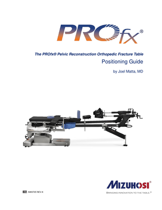
The PROfx® Pelvic Reconstruction Orthopedic Fracture Table
Positioning Guide by Joel Matta, MD
NW0705 REV A
The information contained within this Positioning Guide is not intended to be a substitute for the PROfx® Pelvic Reconstruction Orthopedic Fracture Table Owner’s Manual. Please take the necessary time to read and understand the contents of the Owner’s Manual prior to setting up or operating this table.
Introduction: The PROfx® table is specifically designed to enhance reduction and fixation of acetabular and pelvic fractures. Surgical reduction of these injuries remains a formidable challenge. Benefits of the table can be an easier reduction, making closed reduction possible, making open reduction possible through a lesser approach, and making the most difficult reductions possible at all. The narrow contact area the table makes with the pelvic area also enhances access. For acetabular fractures, the patient is positioned prone for the Kocher-Langenbeck, supine for the ilioinguinal, and laterally for the extended iliofemoral approaches. The table and specific positioning enhances surgical exposure, aids reduction and helps protect the sciatic nerve from stretch injury. The capabilities of the KL and II approaches are maximized and the necessity of utilizing two approaches or the EIF is minimized. For pelvic fractures, the table enhances exposure and aids reduction. The most difficult fracture types are associated with a cranial (vertical) migration of the hemi pelvis. Distal traction applied with the table can be very effective in correcting cranial displacement. However, anterior ring injuries (ramus fracture and symphysis dislocation) can be further displaced if the table’s perineal post is used as counter traction. Table-skeletal fixation to the uninjured side provides the necessary stabilizer (counter traction) against the opposite injured side traction without the disadvantage of the perineal post. If the injury is easily reduced, the counter traction of the patient’s weight may be adequate with the table positioned in maximum Trendelenburg. This booklet illustrates the setup positions I routinely use for acetabular and pelvic fracture surgery. Joel Matta, MD
PROfx® Positioning Guide
3
Figure 1. Acetabular Fractures Prone with Skeletal Traction (Kocher-Langenbeck): Patient is prone on the PROfx® table without table-skeletal fixation with perineal post 6850-413 with a transcondylar distal femoral pin and leg flexed. To facilitate distraction of the femoral head away from the joint, unlock the leg spar and lower leg spar half way down.
Figure 2. Acetabular Fractures Prone with Skeletal Traction (Kocher-Langenbeck): Acetabular fracture reduction and fixation through the KL approach. Patient is prone, with spar lift assist on operative side, distal femoral pin traction, knee flexed 70 degrees, slight internal rotation, ball joints co-axial with traction device and both hips extended. Knee flexion and hip extension on operative side relaxes sciatic nerve to help prevent injury. Opposite side traction lock and slight tension will resist pelvic rotation when traction is applied to injured side. Spar U-joints are just lateral to hip centers.
4
PROfx® Positioning Guide
Figure 3. Ilioinguinal Approach: Level table, place patient supine on the PROfx® table with both arms out to the side, U-joints just lateral to hip center and lower extremities parallel. Pull patient distally against perineal post and center patient on table. Use spar lift assist on operative side and leg supports under both calves. Involved hip is flexed 20 degrees to relax the iliopsoas muscle. Place slight traction tension on patient’s well leg and lock gross traction. Both legs are in neutral rotation.
Figure 4. Ilioinguinal Approach: Unlock traction mount at the ball joint and gross traction to raise lower leg supports until desired flexion is achieved, then lock traction mount ball joint and gross traction.
PROfx® Positioning Guide
5
Figure 5. Supine Fixation of the Symphysis Pubis: Place patient supine on the PROfx® table with both hips equally flexed and adjust ball joints medially or laterally as needed to allow for x-ray views.
Figure 6. Supine Fixation of the Symphysis Pubis: Internal rotation of both legs is used to facilitate reduction.
6
PROfx® Positioning Guide
Figure 7. Prone Position Fixation of Posterior Pelvic Ring: Place patient prone on the PROfx® table with spar ball joints fully abducted using controls at the head end of the table.
Figure 8. Prone Position Fixation of Posterior Pelvic Ring: Place patient’s arms at 90 degrees. If possible, slightly tilt fingers towards the ground. Ensure patient makes firm contact with the perineal post while pulling gross traction on both legs.
PROfx® Positioning Guide
7
Figure 9. Prone Position Fixation of Posterior Pelvic Ring: With patient’s hips extended, raise both leg spars to maintain lower back lordosis and correct position of pelvis. Place lower leg supports under distal thigh and knee cap.
Figure 10. Prone Position Fixation of Posterior Pelvic Ring: Adduct both leg spars using controls at the foot end of the PROfx® leg spars. Prior to prep and drape, check with fluoroscope to make sure proper views, including caudad and cephalad views, can be obtained.
8
PROfx® Positioning Guide
Figure 11. Prone Position Fixation of Posterior Pelvic Ring: Patient is positioned prone on the PROfx® table.
Figure 12. Prone Position Fixation of Posterior Pelvic Ring: Patient is positioned prone on the PROfx® table. The table applies traction and the patient’s weight provides the counter traction. This position is achieved using the Trendelenburg feature on the Hand Pendant.
PROfx® Positioning Guide
9
Figure 13. Pelvic Fracture Skeletal Traction with Operative Leg at 70 Degrees: Patient is in the prone position on the PROfx® table with table-skeletal fixation of the non-operative hip and skeletal traction on the operative side. Distal femoral Steinman pin is attached to a Kirschner tension bow for skeletal traction to operative side. Knee is flexed about 70 degrees. The traction mount ball joint is positioned co-axial with the traction device and the limb is in neutral rotation.
Figure 14. Pelvic Fracture Skeletal Traction with Operative Leg at 70 Degrees: Patient is in the prone position on the PROfx® table with a transcondylar distal femoral pin and leg flexed. NOTE: No perineal post is used.
10
PROfx® Positioning Guide
Figure 15. Posterior Pelvic Ring Injury Reduction and Fixation: Patient is positioned on the PROfx® table in the prone position for posterior pelvic ring injury reduction and fixation. Pelvic frame is placed close to uninjured side. Higher bar for clamp is closer to head of table. Leg spars are extended and supports under knees hyperextend hips to maintain lumbar lordosis. Remove perineal post. Remove central bar for jack attachment. Prior to prepping and draping patient, check to make sure that AP, caudad and cephalad views can be obtained with image intensifier. Possible image problems: patient too proximal or distal and not centered over genital relief hole, leg support post too proximal, arm board support post too distal, or spar U-joints too medial.
Figure 16. Posterior Pelvic Ring Injury Reduction and Fixation: Patient is positioned prone on the PROfx® table with spar ball joints abducted slightly further than patient’s hip width apart using controls at the head end of the table. Prior to prepping and draping patient, place 6 mm external fixator pin into proximal femur at the level of the lesser trochanter of the uninjured side and check with the fluoroscope. Attach pin with ex fix clamps and bars to frame and triangulate bars to increase rigidity. After prepping and draping, place the second 6 mm pin into the posterior superior iliac spine and along the track of the sciatic buttress toward the anterior inferior iliac spine. Attach an ex fix clamp and bar to the pin and pelvic frame by passing the bar through a hole cut in the drapes. An unscrubbed assistant will attach the bar to the frame and pull tension on the bar prior to tightening the clamp. PROfx® Positioning Guide
11
Figure 17. Posterior Pelvic Ring Injury Reduction and Fixation: With patient prone on the PROfx® table, the operative leg spar ball joint should be in line with the traction on the leg and the knee should be flexed at 70 degrees.
12
PROfx® Positioning Guide
Figure 18. Posterior Pelvic Ring Injury Reduction and Fixation: With patient prone on the PROfx® table, the hip needs to be extended to avoid sciatic nerve damage.
PROfx® Positioning Guide
13
Figure 19. Pelvic Fracture Using Table Skeletal Fixation: Patient is positioned prone on the PROfx® table using Mizuho OSI Well Hip Fixation Frame. Attach the Well Hip Fixation Frame and mounting bracket securely to the PROfx® leg spar on the non-operative side. NOTE: Position frame close to patient. Position PRIOR to prepping and draping.
Figure 20. Pelvic Fracture Using Table Skeletal Fixation: Position PRIOR to prepping and draping. Lower femur pin goes into proximal femur at the level of the lesser trochanter. External fixation bars are triangulated to resist distal movement with traction.
14
PROfx® Positioning Guide
Figure 21. Pelvic Fracture Using Table Skeletal Fixation: Position AFTER prepping and draping. Entire top pin enters posterior superior ilium / sciatic buttress.
Figure 22. Pelvic Fracture Using Table Skeletal Fixation: Pin perforates the drape and is placed under tension before locking. It is used to prevent rotation.
PROfx® Positioning Guide
15
Figure 23. Pelvic Fracture Using Table Skeletal Fixation: Additional detail.
Figure 24. Pelvic Fracture Using Table Skeletal Fixation: Additional detail. NOTE: No perineal post is needed for this setup.
16
PROfx® Positioning Guide
Figure 25. Lateral Position for Extended Iliofemoral Approach: Patient positioned laterally on the PROfx® table (front view). Torso support is on sternum. Transverse perineal post is attached to jack and placed posterior to anterior, starting with moderate pressure on medial thigh. Spar lift assist is on the operative side.
Figure 26. Lateral Position for Extended Iliofemoral Approach: Patient positioned laterally on the PROfx® table (rear view).
PROfx® Positioning Guide
17
Figure 27. Lateral Acetabular Extended Iliofemoral Approach: Patient positioned laterally on the PROfx® table. Operative leg spar should be flexed slightly.
Figure 28. Lateral Acetabular Extended Iliofemoral Approach: Patient positioned laterally on the PROfx® table. Operative leg spar should be slightly adducted.
18
PROfx® Positioning Guide
Figure 29. Lateral Acetabular Extended Iliofemoral Approach: Patient positioned laterally on the PROfx® table. Patient’s knee is positioned at 70 degrees with slight internal rotation.
Figure 30. Lateral Acetabular Extended Iliofemoral Approach: Patient positioned laterally on the PROfx® table. Patient’s non-operative hip is extended with slight internal rotation. Traction mount ball joint on operative side is flexed.
PROfx® Positioning Guide
19
Figure 31. Lateral Acetabular Extended Iliofemoral Approach: Patient positioned laterally on the PROfx® table (front view). Patient’s left (non-operative) leg and traction boot are attached to the traction device on the right (operative) side of the table and the patient’s right (operative) leg and traction boot are attached to the prone skeletal traction device on the left (non-operative) side of the table. Skeletal traction is achieved with a transcondylar distal femoral pin and traction device. Both hips are extended and hip extension combined with knee flexion on the operative side relaxes the sciatic nerve to help prevent injury.
Figure 32. Lateral Acetabular Extended Iliofemoral Approach: Patient positioned laterally on the PROfx® table (rear view). Patient’s left (non-operative) leg and traction boot are attached to the traction device on the right (operative) side of the table and the patient’s right (operative) leg and traction boot are attached to the prone skeletal traction device on the left (non-operative) side of the table. Skeletal traction is achieved with a transcondylar distal femoral pin and traction device.
20
PROfx® Positioning Guide