OCULUS
Pentacam Interpretation Guideline Nov 2005
Interpretation Guideline
49 Pages

Preview
Page 1
Pentacam Interpretation Guideline
Copyright by
G/70700/1105/e
Page 2 Pentacam Interpretation Guideline
Foreword We thank you for the trust you have put in this OCULUS product. With the purchase of this instrument, you have chosen a modern, sophisticated product, which was manufactured and tested according to strict quality criteria. Our enterprise has been doing business for over 100 years. Today OCULUS is a medium-sized enterprise concentrating completely on helping ophthalmologists, optometrists and opticians to carry out their responsible work by supplying an optimal range of instruments for examinations and surgery on the eye.
OCULUS has been certified according to DIN EN ISO 9001:2000 and 13485:2003 and therefore sets high quality standards in the development, production, quality assurance and servicing of its entire product range.
The Pentacam is the newest product in the Oculus line. It is based on the Scheimpflug principle, which generates precise, sharp images of the anterior eye segment. Our painstaking product development has produced an instrument that takes extremely accurate measurements and is easy to use. If you have questions or desire further information on this product, call, fax or email us. Our service team will be glad to help you. OCULUS Optikgeräte Managing director and management team
Page 3 Pentacam Interpretation Manual
Introduction This guideline should help all Pentacam users to interpret the results and screens the Pentacam provides. We may not have covered everything which might be of kind of interest. Therefore we ask each Pentacam user for help to improve this guideline step by step. Please forward your special cases to us and we will be happy to implant them.
Of course, this guideline cannot replace the years of experience and the medical studies, but it will be a help in questionable cases as well as be a help for beginners. The personal experience and impression from each of you and the cross connection of the results from different instruments linked with the individual patient’s history may sometimes lead to different results as shown in this guideline.
Page 4 Pentacam Interpretation Guideline
Table of Contents Introduction...3 Table of Contents ...4 1. Description of unit and general remarks...5 2. Corneal INTACS ...6 2.1. Case 1, INTACS after PRK, Alain-Nicolas Gilg, MD...6 3. Orthokeratology ...9 3.1. Case 1, General Screening, Alain-Nicolas Gilg, MD ...9 4. Corneal Ectasia ...12 4.1. Case 1, Ectasia after RK, Renato Ambrósio, MD ...12 4.2. Case 2, Ectasia after LASIK?, Prof. Michael Belin ...14 5. Glaucoma ...17 5.1. Case 2, General screening, Tobias Neuhann, MD ...17 5.2. Case 1, YAG Laser Iridectomy, Eduardo Viteri, MD...18 5.2.1. Comments...20 6. Keratoconus...21 6.1. Case 1, Locating the cone, Prof. Michael Belin ...21 6.2. Keratoconus detection, Prof. Michael Belin ...22 6.2.1. Case 2, Keratoconus, OD & OS?, Prof. Michael Belin ...22 6.2.2. Case 3, INTACS implantation, Prof. Michael Belin...24 6.2.3. Case 4, Form Fruste Keratoconus?, Prof. Michael Belin ...26 6.3. Proposed Screening Parameters, Prof. Michael Belin...28 6.4. Case 5, Unilateral Keratoconus?, Renato Ambrósio, MD...29 6.4.1. Conclusion ...31 7. IOL-calculation after corneal laser refractive surgery...32 7.1. Holladay Report ...32 7.2. Case 1, Tobias Neuhann, MD...34 8. PIOL, pre-op and post-op evaluation, Eduardo Viteri, MD ...35 8.1. Evaluation in Artisan Phakic IOL...35 8.1.1. Preoperative evaluation ...35 8.1.2. Postoperative evaluation ...36 9. Cataract ...37 9.1. Case 1, Cortical Cataract, Tobias Neuhann, MD...37 10. Corneal transplant ...38 10.1. Case 1, Removing the sutures?, Tobias Neuhann, MD...38 11. What would you recommend? ...39 11.1. Case 1, Keratoconus and Cataract, Tobias Neuhann, MD...39 12. Other cases ...42 12.1. Case 1, Corneal Infiltrate, Renato Ambrósio, MD ...42 12.2. Case 2, Incisional Edema, Renato Ambrósio, MD ...43 12.3. Case 3, Corneal Thinning after Herpetic Keratitis, Renato Ambrósio, MD ...44 12.4. Case 4, Epithelial Ingrowth after Keratomileusis in situ, Renato Ambrósio, MD ...45 13. Recommended Settings and Color Maps...46 13.1. Recommended Settings ...46 13.2. Recommended Color Maps...46 13.2.1. Screening for LASIK, PRK etc. ...46 13.2.2. Screening for PIOL implantation ...47 13.2.3. Glaucoma Screening...47 13.2.4. IOL Calculation for Treated and Untreated Corneas ...47 13.2.5. Screening for Keratoconus and Ectasia...48 14. References and Contact Addresses...49
Page 5 Pentacam Interpretation Manual
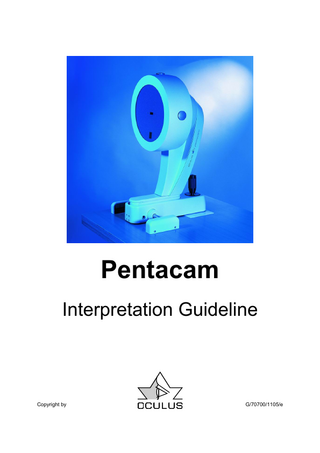
1. Description of unit and general remarks The OCULUS Pentacam is a rotating Scheimpflug camera. The rotational measuring procedure generates Scheimpflug images in three dimensions, with the dot matrix finemeshed in the center due to the rotation. It takes a maximum of 2 seconds to generate a complete image of the anterior eye segment. Any eye movement is detected by a second camera and corrected for in the process. The Pentacam calculates a 3-dimensional model of the anterior eye segment from as many as 25,000 true elevation points. The topography and pachymetry of the entire anterior and posterior surface of the cornea from limbus to limbus are calculated and depicted. The analysis of the anterior eye segment includes a calculation of the chamber angle, chamber volume and chamber height and a manual measuring function at any location in the anterior chamber of the eye. In a moveable virtual eye, images of the anterior and posterior surface of the cornea, the iris and the anterior and posterior surface of the lens are generated. The densitometry of the lens is automatically quantified. The Scheimpflug images taken during the examination are digitalized in the main unit and all image data are transferred to the PC. When the examination is finished, the PC calculates a 3D virtual model of the anterior eye
segment, from which all additional information is derived. OCULUS Optikgeräte GmbH emphasizes that the user bears the full responsibility for the correctness of data measured, calculated or displayed using the Pentacam. The manufacturer will not accept claims based on erroneous data and wrong interpretation. This interpretation guideline has to be understood as a help only to interpret the examination data the Pentacam provides. The doctors and physicians have to consider all medical information which can be collected by using other diagnostic instruments e.g. slit lamp examination, ultrasound biomicroscopy, etc. to make the diagnosis. The results of the different diagnostic instruments have to be compared and closely scrutinized. This interpretation guideline has to be understood as a completion to the operator’s manual. The current version of the operator’s manual is stored on every Pentacam software CD-ROM.
Page 6 Pentacam Interpretation Guideline
2. Corneal INTACS 2.1. Case 1, INTACS after PRK, Alain-Nicolas Gilg, MD A female, 45 years-old, had PRK in both eyes 7 years before. The visual acuity before the laser surgery was • OD sph. -7.50, cyl. -0.50 @170°, • OS sph. -6.75, cyl. -1.00 @10°.
Figure 1, Zernike Analysis
She was referred for blurred vision, photophobia, and poor intermediate VA. The Zernike analysis confirmed the functional disorders of the vision due to abnormal spherical and high order aberrations, |Z|40 (spherical), |Z|53.(trefoil 5th order) |Z|62 (astigmatism 6th order) (Figure 1).
Page 7 Pentacam Interpretation Manual
The Keratoconus menu of the Pentacam identifies this cornea as an oblate postoperative cornea, note the negative eccentricity and display an abnormal high aberration coefficient due to the high order aberrations (Figure 2). The Pachymetry map shows a smooth progression with a thick area for the implantation of the
Figure 2, Keratoconus Menu
INTACS in the 7mm zone. Though, she was a good candidate for INTACS implantation. The visual acuity before the implantation of the corneal INTACS was: • OD 0.9; sph. -1.25, cyl. -0.50 @175° • OS 0.6; sph. -1.50, cyl. -0.50 @55°.
Page 8 Pentacam Interpretation Guideline
The visual acuity after implantation of the INTACS: • OS 1.0; sph. +0.50, cyl. -1.25 @30°.
Figure 3, Scheimpflug Image
The Scheimpflug image shows a successful fit of the implanted INTACS (Figure 3).
Page 9 Pentacam Interpretation Manual
3. Orthokeratology 3.1. Case 1, General Screening, Alain-Nicolas Gilg, MD A male, 34 years old, referred for changing his soft contact lenses because of a progressive intolerance during the day. The subjective refraction results in visual acuity • OD sph. -2.5, • OS sph. -1.0.
Figure 1, Show 2 Examinations
The Pentacam “Show 2 examinations screen” displays an optimal eccentricity on the 30° of both eyes, OD 0.50, OS 0.49 which permits us to propose the orthokeratology treatment to this patient (Figure 1). After fitting the lenses, prior to midday examinations revealed a good visual acuity on day 1, VA: 0.8, day 8 and 28 VA: 1.0.
Page 10 Pentacam Interpretation Guideline
The patient was examined 4 times within 2 months to view the corneal progression. The 4
maps comparison screen confirmed the efficacy of the treatment (Figure 2).
Figure 2, 4 Maps Comparison On day 28, the patient complained of fluctuations during the day of his visual acuity. The Patient was examined in the morning after wearing the lens over night and in the late afternoon. The Pentacam confirmed using the “Compare 2
Figure 3, 2 Maps Comparison
Exams” screen that the effect of the ortho-K lens was reversible during the day which leads to the diagnosis to fit a more effective ortho-K lens to this patient (Figure 3).
Page 11 Pentacam Interpretation Manual
This page lefts international free.
Page 12 Pentacam Interpretation Guideline
4. Corneal Ectasia 4.1. Case 1, Ectasia after RK, Renato Ambrósio, MD A 28 years old male patient had RK in 1995 for myopic astigmatism with RK enhancement three years later in OS. Corneal topography was not performed prior to surgery according to patient information. Uncorrected VA was 20/30 in OD and 20/200 in OS. Patient refers severe glare and starburst all day, mainly at night. Refraction is –0.25 –3.00 x 156, giving 20/20 in
Figure 1, 4 Maps Refractive
OD and –5.00 –2.25 x 39, giving 20/30 in OS. Patient was fit with a RGPCL with significant improvement of the symptoms in both eyes. The Pentacam Quad map demonstrates corneal Ectasia in both eyes, more advanced in OS (Figure 1) In OD, (Figure 2) the patient has a central cornea with less distortion than OS, which enables relatively good uncorrected vision. However, the patient refers quality of vision was terrible in both eyes.
Page 13 Pentacam Interpretation Manual
Figure 2, 4 Maps Refractive
Figure 3, Pachymetry Progression
Figure 4, Pachymetry Progression
The pachymetric progression is abrupt in both eyes as an important sign of Ectasia (Figure 3, 4). Probably mild Ectasia could have been diagnosed prior to surgery if corneal topography
and tomography would have performed and well interpreted. This case would have been considered as a bad candidate for RK.
Page 14 Pentacam Interpretation Guideline
4.2. Case 2, Ectasia after LASIK?, Prof. Michael Belin A 46 year old female had previous LASIK 2 years prior. She presented interested in an enhancement to her dominant right eye. BSCVA was 20/20+ with – 1.25 D.
Figure 1, ORB-Scan 4 Maps
The referring surgeon was concerned about Post LASIK Ectasia based on OrbScan topography. OrbScan topography shows significant posterior elevation (Figure 1).
Page 15 Pentacam Interpretation Manual
Evaluation with the OCULUS Pentacam reveals no posterior elevation abnormality and no evidence of post-operative Ectasia (Figure 2).
Patient underwent a routine LASIK enhancement without incident.
Figure 2, 4 Maps Refractive DISCUSSION - This case demonstrates one of the limitations with the current version of the B&L OrbScan. The OrbScan routinely fails to correctly identify the posterior corneal surface in postoperative patients leading to underestimates of residual bed thickness and frequent incorrect diagnosis of Post LASIK Ectasia.
Here the OrbScan incorrectly reads the corneal thickness 37µm thinner than the Pentacam and shows an incorrect Ectasia (Figure 3). The Pentacam shows a normal post-operative appearance (Figure 3).
Page 16 Pentacam Interpretation Guideline
Orbscan 37 µm thinner Figure 3, ORB-Scan 4 Maps, Pentacam 4 Maps Refractive
Page 17 Pentacam Interpretation Manual
5. Glaucoma 5.1. Case 2, General screening, Tobias Neuhann, MD A 48 year old white male patient wants to have a second opinion about his glaucoma treatment. His father and grandfather have had glaucoma. He himself has had ten years of glaucoma
medical treatment. His ophthalmologist recommends now a second medication. We measured 24mmHG with Goldmann applanation tonometer.
Figure 1 , 4 Maps Refractive After taking a Pentacam examination, looking to the 4 maps display (Figure 1) we put the 24mmHg in the Dresdner scale and the corrected IOD was displayed with 11mmHg because of a corneal thickness of about 728µm in the apex. The additional examination on HRT
resulted in a healthy optic nerve and we recommend the patient to stop his medication. His IOP today is during daytime between 19 and 22mmHg. We still see him 4 times a year for IOD and HRT check (Figure 2, 3).
Figure 2, HRT Image
Figure 3, HRT Image
Page 18 Pentacam Interpretation Guideline
5.2. Case 1, YAG Laser Iridectomy, Eduardo Viteri, MD This is a 64 year old female patient who was complaining of episodes of blurred vision and tearing. The IOP was 18 mm Hg in both eyes. Anterior chamber was shallow on slit lamp examination and optic nerve had a C/D ratio of 0.6 in both eyes. The lens was clear and gonioscopy exam revealed a narrow angle in both eyes (grade I-II).
Figure 1, Overview pre-op
The anterior segment exam with the Pentacam (Figure 1) documented an irido-corneal angle of 22.5 degrees with an ACD (epithelial) of 2.43 mm. The patient was reluctant to have YAG laser Iridectomy until she was able to compare her anterior segment biometry with that of other normal patients.
Page 19 Pentacam Interpretation Manual
After YAG Laser Iridectomy was performed, several of her anterior segment measurements
Figure 2, Overview post-op
changed (Figure 2). This is quite evident in the differential display (Figure 3)
Page 20 Pentacam Interpretation Guideline
Figure 3, 2 Maps Comparison The irido-corneal angle is 4º wider, and, although the ACD only deepened 0.09 mm centrally, the main difference is evident in the periphery,
5.2.1. In narrow angle Glaucoma, the Pentacam is quite useful in measuring the irido-corneal angle, although this may be difficult in 360º because of the eyelid interference. We can obtain more consistent data when measuring peripheral ACD
where you can see changes ranging from 0.19 mm to 0.30 mm. This was enough to increase the AC volume from 64 to 92 mm3.
Comments and AC volume. The exam has been of great help also in educating the patient about this disease, and making evident the effect of the treatment.