PULSION Medical Systems
PiCCO2 Information for Nurses May 2010
Guide
33 Pages

Preview
Page 1
Get the complete picture ...
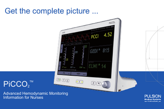
PiCCO2
TM
Advanced Hemodynamic Monitoring Information for Nurses
PiCCO2 - A complete hemodynamic picture TM
• Continuous cardiac output • Volumetric preload • Afterload • Contractility • Volume responsiveness • Pulmonary edema / Lung water
PiCCO2TM 2
Table of contents Overview...
4-6
Fields of Application ...
7
PiCCO2TM Disposables and Set up ...
8 - 11
Principles of PiCCO2TM Technology... 12 - 14 Parameter Overview... 15 - 25 PiCCO2TM Monitor... 26 Normal Ranges... 27 Decision Tree Model... 28 Summary of Benefits... 29 Recommended Literature... 30 Contact Information... 32
3
Cardiac Output - Optimizing Oxygen delivery
O2 uptake
O2 transport
O2 extraction
O2 utilization
PiCCO-Technology Which therapy?
Volume?
Vasopressors?
Inotropes? 4
Is measuring just the CO enough? Cardiac output CO Stroke volume SV
Heart rate HR
Preload GEDV, SVV, PPV
Afterload SVR, MAP
Contractility CFI
Pulmonary edema EVLW
Volume?
Vasopressors?
Inotropes? 5
PiCCO2 – See more than others TM
Optimize CO
Protect the lungs
• Continuous cardiac output
• Monitor Pulmonary Edema (lung water) at the bedside.
• Volumetric preload • Afterload • Contractility
• Respond quickly • More reliable than Chest X Ray
• Volume responsiveness
6
Fields of Application Intensive Care
OR/Post surgery
• Septic Shock
• Cardiac Surgery
• Cardiogenic Shock
• Major Surgery
• Burns
• Neuro Surgery
• Trauma / Hypovolemic Shock
• Pediatrics
• ARDS / Acute Lung Injury • Pediatrics
7
Use your existing CVC and the PiCCO arterial line
Central venous line
(Standard CVC) Internal jugular, subclavian, femoral
Arterial line
(PiCCO Catheter available in different sizes) Brachial, axillary or femoral artery
8
PiCCO arterial catheters - The choice is yours Axillary artery Adults: 4F 8 cm / 3.15 in Small adults: 3F 7 cm / 2.76 in
Brachial artery Adults: 4F 16 cm / 6.29 in Adults: 4F 22 cm / 8.66 in
Femoral artery Adults: 5F 20 cm / 7.78 in Adults: 4F 22 cm / 8.66 in Small adults: 4F 16 cm / 6.29 in Children: 3F 7 cm / 2.76 in and 4F 8 cm / 3.15 in 9
PiCCO2 Setup TM
Thermodilution
C B CVP line
Flush bag
A
PiCCO Catheter
B
Distal lumen of CVC
C
Injectate sensor housing
D
Injectate sensor cable
E
Arterial connection cable
F
PiCCO thermistor plug Pressure connection cable
G
D
F
E
Pressure output adapter A
G 10
PiCCO - Two principles for reliable information Transpulmonary thermodilution
Pulse contour analysis
Calibration
Intermittent parameters from thermodilution technique • Thermodilution cardiac output (CO) / cardiac index (CI) • Volumetric preload (GEDV/GEDI) • Contractility (CFI) • Lung water (EVLW/ELWI)
Continuous parameters from analysis of the arterial waveform (pulse contour) • Pulse contour cardiac output (PCCO) / cardiac index (PCCI) • Afterload (SVR/SVRI) • Volume responsiveness (SVV, PPV) • Stroke volume (SV/SVI) 11
PiCCO - Transpulmonary Thermodilution (TD) Bolus injection
Bolus detection
• The bolus injection of cold saline is detected by the temperature sensor attached to the CVC • The saline bolus passes through the heart and lungs and is detected downstream by the thermistor on the tip of the PiCCO arterial line • The change in temperature between the two thermistors provides a TD curve • Mathematical analysis of the TD curve gives the cardiac output CO • Further analysis of the TD curve allows determination of preload volumes and lung water 12
PiCCO - Pulse Contour Analysis
• Stroke volume is the area under the systolic part of the pressure curve (red area) of one heart beat • Cardiac output is calculated and updated beat-by-beat: stroke volume x heart rate • In severely “shocked” patients organ perfusion pressure and pulse contour CO is more reliably represented by central arterial pressure (femoral / brachial / axillary) than by radial arterial pressure 13
CCI
Thermodilution (TD) Cardiac Output / Index Cardiac Output - Volume of blood pumped by the heart in one minute - Important determinant for oxygen delivery to the body
Time since TD 0 h 52 min 10:26 am TD Results
10:18 am 10:20 am 10:22 am 10:26 am SEP 23 SEP 23 SEP 23 SEP 23
CI
3.47
3.11
3.15
3.76
3.44
GEDI
705
626
678
764
698
ELWI
9
9
10
10
9
T
1.18
1.17
0.30
1.20
Flow
Inj. Volume 98.1
START READY
10:26 am SEP 23
15 ml CVP 5
98.6 0s
10s
20s
30s
mmHg
PCCI
5.0 3.0
Exit
SVRI 1735
dyn*s*cm-5 m 2
4.52 l/min/m 2
SVI
ml/m 2
47
CO – Cardiac Output CI – Cardiac Index The Thermodilution CO is used to “calibrate“ the continuous CO obtained from 2the arterial waveform l/min/m (pulse contour) analysis. Volume
14
s
Pulse Contour Cardiac Output / Index Cardiac Output - Following thermodilution a “calibrated” continuous CO can be obtained Time since TD 0 h 52 min 10:26 am Flow
PCCI
PCCI
SVRI 1735
5.0 3.0
dyn*s*cm-5 m 2
PCCO – Pulse Contour Cardiac Output PCCI – Pulse Contour Cardiac Index Volume • Product of stroke volume and heart rate • Determination beat-by-beat MAP • Reliability and patient safety possible due to calibration technique
4.52 l/min/m 2
SVI
ml/m 2
47
l/min/m 2
ml/m 2
15
l/min/m 2
SVRIpressures 1735 Preload Volume instead of filling PCCI dyn*s*cm m -5
2
SVI
ml/m 2
47
Preload - Volume of blood in the heart, available to be pumped Volumetric preload parameters are superior to filling pressures (CVP / PCWP) Michard, YICM 2004 l/min/m 2
Volume
MAP ml/m 2
SVV
9 %
GEDV – Global End-Diastolic Volume / GEDI – Global Volume Index OrganEnd-Diastolic Function • Combined diastolic volume of all four heart chambers • Adequate preload is an important prerequisite for adequate cardiac output (Frank-Starling curve) • GEDI is indexed to “predicted body surface area” *
SVRI
* Indexing particular parameters i.g. to the predicted body weight (EVLW) or predicted body surface area (GEDV) rather than the actual body weight or body surface area is more accurate particularly in overweight patients. ml/kg 16
Volume
MAP edema assessment at the bedside Lung Water – Pulmonary EVLW – Extravascular Lung Water reflects pulmonary edema
SVV
ml/m 2
9 %
Organ Function
SVRI ml/kg
AP/CVP 1/minIndex EVLW – Extravascular Lung Water / ELWI – Extravascular Lung Water • Extravascular lung water (EVLW) represents the extravascular water content of the lung tissue • Includes intra-cellular, interstitial and intra-alveolar water (not pleural effusion) • ELWI is indexed to “Predicted Body Weight” *Predicted body weight is determined from the body height, gender and age 17
Fluid management using lung water (ELWI) Improved patient outcomes
Organ function - Lung water Ventilation days
Comparison of fluid therapy targeting lung water (protocol group) or wedge pressure (standard group)
ICU days
reduced by
reduced by
Days
59%
Control group PCWP
Protocol group EVLW
53%
Control group PCWP
Protocol group EVLW
Source: Improved outcome based on fluid management in critically ill patients requiring pulmonary artery catheterization Mitchell JP, Schuller D, Calandrino FS, Schuster DP, Am Rev Respir Dis 1992; 145(5): 990-8 18
Time since TD 0 h 52 min 10:26 am Flow resistance Afterload - the systemic vascular
PCCI
5.0
4.52
Systemic vascular resistance - Represents the vascular “tone” of the blood3.0 vessels.
SVRI 1735
PCCI
dyn*s*cm-5 m 2
l/min/m 2
SVI
ml/m 2
47
l/min/m 2 Volume
MAP
SVR - Systemic Vascular Resistance SVRI - Systemic Vascular Resistance Index • Helps to determine levels of vasodilation or vasoconstriction • Useful for guiding vasopressor management.
ml/m 2 19
ml/m 2
SVV
Heart Contractility
9
% Contractility – describes the performance of the cardiac muscle Organ Function
SVRI ml/kg
AP/CVP
1/min
CFI - Cardiac Function Index • Parameter of the global cardiac contractility • The cardiac function index is the ratio of flow and preload • CFI = CO (Cardiac Output) / GEDV (Global End-Diastolic Volume) 20