PULSION Medical Systems
PiCCOplus Principles of Operation July 2003
Guide
27 Pages

Preview
Page 1
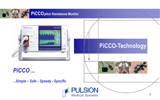
PiCCO plus Standalone Monitor
PiCCO-Technology
PiCCO ... ...Simple – Safe – Speedy - Specific
1
Contents 1. What is the PiCCO-Technology?... 3 2. What are the advantages of the PiCCO-Technology?... 4
3. How does the PiCCO-Technology work?... 6 4. How to use the PiCCO-Technology?... 19 5. Which disposables do I need for the PiCCO-Technology?... 22 6. References…………………………………………………………………………………….. 24 7. Where can I get what I need?... 25
2
1. What is the PiCCO-Technology?
The PiCCO Technology is a combination of 2 techniques for advanced hemodynamic and volumetric management without the necessity of a pulmonary artery catheter in most patients: a. Transpulmonary thermodilution
b. Arterial pulse contour analysis
-∆T
-∆T
see also page 8
t
see also page 16
t
3
2. What are the advantages of the PiCCO-Technology?
The PiCCO measures the following main parameters: Thermodilution Parameters • Cardiac Output • Global Enddiastolic Volume • Intrathoracic Blood Volume • Extravascular Lung Water
CO GEDV ITBV EVLW*
Pulse Contour Parameters • Pulse Continuous Cardiac Output PCCO • Systemic Vascular Resistance SVR • Stroke Volume Variation SVV * not available in USA
4
2. What are the advantages of the PiCCO-Technology? Less Invasiveness
- Only central venous and arterial access required - No pulmonary artery catheter required - Applicable also in small children
Short Set-up Time
- Can be installed within minutes
Dynamic, Continuous Measurement - Cardiac Output, Afterload and Volume Responsiveness are measured beat-by-beat No Chest X-ray
- To confirm correct catheter position no x-ray is necessary
Cost Effective
- Less expensive than pulmonary artery catheter technique - Arterial PiCCO catheter can be in place for 10 days or more - Potential to reduce ICU stay and costs
More Specific Parameters
- PiCCO parameters are easy to use and interpret even for less experienced clinical staff
Extravascular Lung Water*
* not available USA - Lung edema can be excluded or quantified at thein bed-side 5
3. How does the PiCCO-Technology work? Most of hemodynamic unstable and/or severely hypoxemic patients are instrumented with:
Central venous line Arterial line
(e.g. for vasoactive agents administration…)
(accurate monitoring of arterial pressure, blood samples…)
The PiCCO-Technology uses any standard CV-line and a thermistor-tipped arterial PiCCO-catheter instead of the standard arterial line.
6
Configuration
Central venous line (CV)
CV
A Thermodilution catheter with lumen for arterial pressure measurement • Axillary (A) • Brachial (B) • Femoral (F) • Radial (R), long catheter
B
R
F
Arterial pressure transducer 7
a. Transpulmonary Thermodilution Transpulmonary thermodilution measurement simply requires the central venous injection of a cold (< 8°C) or room-tempered (< 24°C) saline bolus…
CV Bolus Injection
Lungs Right Heart
Left Heart PiCCO Catheter e.g. in femoral artery 8
PiCCO Thermodilution Cardiac Output After central venous injection of the indicator, the thermistor in the tip of the arterial catheter measures the downstream temperature changes The cardiac output is calculated by analysis of the thermodilution curve using a modified Stewart-Hamilton algorithm: -Tb Injection
t 9
PiCCO Volumetric Parameters Global Enddiastolic Volume GEDV Intrathoracic Blood Volume ITBV Extravascular Lung Water EVLW* These volumetric parameters are obtained by advanced analysis of the thermodilution curve. (Detailed information and formulas available on request.)
Advanced Thermodilution Curve Analysis Tb
injection recirculation
ln Tb
e -1 At
MTt
DSt
t
* not available in USA 10
Global Enddiastolic Volume Global Enddiastolic Volume (GEDV) is the volume of blood contained in the 4 chambers of the heart.
11
Intrathoracic Blood Volume Intrathoracic Blood Volume (ITBV) is the volume of the 4 chambers of the heart + the blood volume in the pulmonary vessels.
12
Extravascular Lung Water* Extravascular Lung Water (EVLW)* is the amount of water content in the lungs. It allows bedside quantification of the degree of pulmonary edema.
* not available in USA 13
PiCCO Preload Indicators Intrathoracic Blood Volume, ITBV and Global Enddiastolic Volume, GEDV have shown to be far more sensitive and specific to cardiac preload than the standard cardiac filling pressures CVP + PCWP but also than right ventricular enddiastolic volume. 2,3,5,6,8,9,12,13,22
The striking advantage of ITBV and GEDV is that they are not wrongly influenced by mechanical ventilation and give correct information on the preload status under any condition. 2,3,6,7,8,9,12,13, 22
14
Extravascular Lung Water* Extravascular Lung Water, EVLW* assessment by transpulmonary thermodilution has been validated against dye dilution and the reference gravimetric method. 11,16,21,23
Extravascular Lung Water, EVLW* has shown to have a clear correlation to severity of ARDS, length of ventilation days, ICU-Stay and Mortality and to be superior to assessment of lung edema by chest x-ray.7,8,15,20,23,24
* not available in USA 15
b. Arterial Pulse Contour Analysis Arterial pulse contour analysis provides continuous beat-by-beat parameters obtained from the shape of the arterial pressure wave. The algorithm is capable of computing each single stroke volume (SV) after being calibrated by an initial transpulmonary thermodilution. -∆T
-∆T
t
P [mm Hg]
t
Calibration SV
t [s]
16
Cardiac Output and Systemic Vascular Resistances As pulse contour analysis continuously measures stroke volume and arterial pressure, cardiac output (CO) and systemic vascular resistance (SVR) are computed as follows: CO is calculated as stroke volume x heart rate SVR is calculated as (mean arterial pressure - central venous pressure) / CO
17
Stroke Volume Variation (SVV) In mechanically ventilated patients without arrhythmia, SVV reflects the sensitivity of the heart to the cyclic changes in cardiac preload induced by mechanical ventilation.1,14,17,18,19 SVV can predict whether stroke volume will increase with volume expansion.1,14,17,18,19
18
4. How to use the PiCCO-Technology? 1. Connect the injectate-temperature sensor housing to the CV line already in place. 2. Insert a PiCCO arterial thermistor catheter into a large artery, preferable femoral artery, but also brachial / axillary artery and radial artery (with long catheter). 3. Connect the injectate sensor, the arterial catheter’s thermistor and pressure line to your PiCCO monitor.
4. For blood pressure transfer to any bedside monitoring system, connect the cable at the back side of the PiCCO monitor. 5. Now the system is ready to work.
6. For information how to handle your PiCCO monitor, please refer to your accompanying PiCCO Operator’s Manual. 19
How to manage my patient with the PiCCO-Technology? Management of a patients hemodynamic situation is easily possible by following the therapeutic guideline shown below.+ It was developed out of daily clinical practice, has shown to be successful in over a hundred thousand patients and refers to below listed normal values of indices: Cardiac Index Global Enddiastolic Blood Volume Index Intrathoracic Blood Volume Index Stroke Volume Variation Extravascular Lung Water Index*
+without guarantee
CI GEDI ITBI SVV ELWI*
3.0 – 5.0 680 – 800 850 – 1000 10 3.0 – 7.0
l/min/m2 ml/m2 ml/m2 % ml/kg
* not available in USA
20