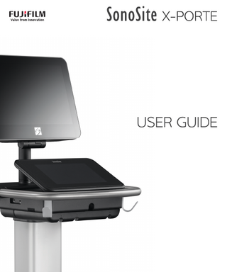
SonoSite
X-Porte User Guide
User Guide
410 Pages

Preview
Page 1
USER GUIDE
Manufacturer
EC Authorized Representative
Australia Sponsor
FUJIFILM SonoSite, Inc.
FUJIFILM SonoSite B.V.
FUJIFILM SonoSite Australasia Pty Ltd
21919 30th Drive SE
Joop Geesinkweg 140
114 Old Pittwater Road
Bothell, WA 98021 USA
1114 AB Amsterdam,
BROOKVALE, NSW, 2100
T: 1-888-482-9449 or 1-425-951-1200
The Netherlands
Australia
F: 1-425-951-1201
Caution
Federal (United States) law restricts this device to sale by or on the order of a physician.
SonoMB, SonoSite Synchronicity, SonoSite, Steep Needle Profiling, X-Porte, and the SonoSite logo are registered and unregistered trademarks of FUJIFILM SonoSite, Inc. in various jurisdictions. FUJIFILM is a registered trademark of FUJIFILM Corporation. Value from Innovation is a trademark of FUJIFILM Holdings America Corporation. DICOM is a registered trademark of the National Electrical Manufacturers Association. All other trademarks are the property of their respective owners. Patents: US 9,895,133; US 9,848,851; US 9,671,491; US 9,420,998; US 9,151,832; US 8,876,719; US 8,861,822; US D712,540; US D712,539; US D712,038; US D712,037; US 8,568,319; US 8,500,647; US 8,398,408; US 8,213,467; US 8,088,071; US 8,066,642; US D625,015; US D625,014; US 7,740,586; US 7,591,786; US 7,588,541; US 6,471,651; US 6,364,839; CA 2,372,152; CA 2,371,711; CN ZL201180028132.X; CN ZL2010800609363; EP 2498683 validated in DE and FR; EP 1589878 validated in DE, FR, and GB; EP 1552792 validated in DE, FR, and GB; EP 1180971 validated in DE and GB; EP 1180970 validated in DE, FR, and GB; JP 6258367; JP 6227724; JP 5973349; JP 5972258. Part Number: P14645-07 Publication Date: June 2019 Copyright © 2019 FUJIFILM SonoSite, Inc. All Rights reserved.
CONTENTS
1. Introduction About the SonoSite X-Porte User Guide ... 1-1 Changes in this version ... 1-2 Document conventions ... 1-2 Getting help ... 1-3
2. Getting started About the system ... 2-1 Intended use ... 2-2 Indications for use ... 2-2 Contraindications ...2-14 Hardware features ...2-15 Accessories and peripherals ...2-16 Preparing the system ...2-16 Turning on the system ...2-16 Adjusting the height and angle ...2-17 USB devices ...2-18 General interaction ...2-19 Clinical monitor ...2-19 VGA or digital video output ...2-21 Touch panel ...2-21 Onscreen keyboard ...2-23 Preparing transducers ...2-24 Connecting transducers ...2-24 Selecting a transducer and exam type ...2-27 Gel ...2-32 Sheaths ...2-33 Ports ...2-33 Battery charge indicator ...2-35 Transporting the system ...2-35 Visual Guide videos ...2-37
3. Setting up the system Administration settings ... 3-1 About security settings ... 3-2 Managing the Administrator account ... 3-2
i
Protecting patient information ... 3-3 Adding and managing user accounts ... 3-3 Configuring Auto Delete ... 3-5 Logging in ... 3-6 Audio settings ... 3-6 Calculations settings ... 3-7 Cardiac calculations settings ... 3-7 Obstetrics calculations settings ... 3-7 CDA Report settings ... 3-11 Connectivity settings ... 3-12 Importing and exporting connectivity settings ... 3-13 DICOM ... 3-14 Configuring the system for DICOM transfer ... 3-15 Connecting to the network ... 3-15 DICOM configuration pages ... 3-17 Associating devices with locations ... 3-26 Date and Time settings ... 3-27 Display Information settings ... 3-28 Logs ... 3-28 Network Status settings ... 3-30 Power and Battery settings ... 3-30 Presets settings ... 3-31 General preferences ... 3-31 Brightness ... 3-32 Labels ... 3-32 Exam types ... 3-33 User profile settings ... 3-34 Importing and exporting ... 3-36 Routing selections ... 3-36 Associating routing selections with exams ... 3-37 Specifying educational DICOM archivers ... 3-38 System Information settings ... 3-38 USB settings ... 3-39 Limitations of JPEG format ... 3-40
4. Imaging Imaging modes ... 4-1 2D ... 4-2 M Mode ... 4-2 Color ... 4-3 Doppler ... 4-4 Dual ... 4-5 Simultaneous Doppler ... 4-7 Imaging controls ... 4-8 Controls in 2D ... 4-9 Controls in M Mode ... 4-14
ii
Controls in Color ... 4-16 Controls in Doppler ... 4-18 Adjusting depth and gain ... 4-22 Depth ... 4-22 Gain ... 4-23 Freezing, viewing frames, and zooming ... 4-24 Freezing the image ... 4-24 Viewing the cine buffer ... 4-24 Zooming in on the image ... 4-25 Visualizing needles ... 4-26 Needle size and angle ... 4-29 Additional recommendations ... 4-29 Labeling images ... 4-30 Adding text labels ... 4-30 Adding arrows ... 4-31 Adding pictographs ... 4-31 Setting the home position ... 4-32 Labeling during review ... 4-32 Entering patient information ... 4-33 Editing patient information ... 4-33 Entering patient information manually ... 4-34 Entering patient information from the worklist ... 4-34 Ending the exam ... 4-37 Patient form fields ... 4-37 Images and clips ... 4-40 Reviewing ... 4-40 Printing images ... 4-43 Archiving and exporting ... 4-44 Saving images and video clips ... 4-47 DVR recording ... 4-49 Image Gallery ... 4-51 ECG ... 4-52
5. Measurements and calculations Measuring ... 5-1 Calipers ... 5-1 Viewing and deleting measurement results ... 5-2 Basic measurements in 2D ... 5-2 Basic measurements in M Mode ... 5-3 Basic measurements in Doppler ... 5-4 Assigning measurements to calculations ... 5-8 About calculations ... 5-8 Overview ... 5-9 Percent reduction calculations ... 5-11 Volume calculation ... 5-12 Volume flow calculation ... 5-12
iii
Exam-based calculations ... 5-13 Abdominal calculations ... 5-13 Arterial calculations ... 5-14 Cardiac calculations ... 5-18 Gynecological calculations ... 5-34 Obstetrics calculations ... 5-37 Small Parts and MSK calculations ... 5-43 Acute Care calculations ... 5-44 Transcranial Doppler and Orbital calculations ... 5-47 Worksheets and reports ... 5-50 Report preview ... 5-51 Acute Care and MSK worksheets ... 5-53 Printing reports and worksheets ... 5-54 Displaying reports after the exam has ended ... 5-54 Customizing worksheets ... 5-55 Remote worksheets ... 5-55 Measuring during review ... 5-57
6. Measurement references Measurement accuracy ... 6-1 Sources of measurement errors ... 6-3 Measurement publications and terminology ... 6-3 Cardiac references ... 6-4 Obstetrical references ... 6-15 General references ... 6-21
7. Troubleshooting and maintenance Troubleshooting ... 7-1 Software licensing ... 7-3 Maintenance ... 7-5 System backups ... 7-5 Servicing ... 7-6
8. Cleaning and disinfecting Before getting started ... 8-1 Determining the required cleaning and disinfecting level ... 8-2 Spaulding classifications ... 8-2 Clean and disinfect system and transducer to a high-level (semi-critical uses) ... 8-3 Clean and disinfect system and transducer to a low-level (non-critical uses)... 8-8 Storing the transducer ... 8-11 Transporting the transducer ... 8-11 Accessories... 8-13
iv
Cleaning and disinfecting accessories ... 8-13 Cleaning and disinfecting the stand or Triple Transducer Connect (TTC) ... 8-14 Cleaning the footswitch ... 8-14 Cleaning and disinfecting the ECG cable and slave cable ... 8-15
9. Safety Ergonomic safety ... 9-1 Position the system ... 9-2 Position yourself ... 9-2 Take breaks, exercise, and vary activities ... 9-3 Electrical safety ... 9-3 Electrical safety classification ... 9-5 Isolating the SonoSite X-Porte ultrasound system from power ... 9-6 Equipment safety ... 9-7 Clinical Safety ... 9-8 Electromagnetic compatibility ... 9-10 Wireless transmission ... 9-12 Electrostatic discharge ... 9-13 Separation distance ... 9-14 Compatible accessories and peripherals ... 9-15 Manufacturer’s declaration ... 9-18 Labeling symbols ... 9-24 Specifications ... 9-33 Dimensions ... 9-33 Environmental limits ... 9-34 Electrical ... 9-34 Imaging modes ... 9-34 Image and video clip storage capacity ... 9-34 Standards ... 9-35 Electromechanical safety standards ... 9-35 EMC standards classification ... 9-36 DICOM standard ... 9-36 HIPAA standard ... 9-36
10. Acoustic output ALARA principle ... 10-1 Applying the ALARA principle ... 10-1 Direct, indirect, and receiver controls ... 10-2 Acoustic artifacts ... 10-3 Guidelines for reducing MI and TI ... 10-3 Output display ... 10-5 MI and TI output display accuracy ... 10-7 Factors that contribute to display uncertainty ... 10-7 Related guidance documents ... 10-8
v
Transducer surface temperature rise ... 10-8 Acoustic output measurement ... 10-9 Tissue models and equipment survey ... 10-10 Acoustic output tables ... 10-11 Acoustic measurement precision and uncertainty ... 10-89 Terminology in acoustic output tables ... 10-90
11. IT Network Functions ... 11-1 Network for connecting the device ... 11-1 Specifications for the connection ... 11-1 Hardware specification ... 11-1 Security ... 11-1 Data flow ... 11-2 IT network failure recovery measures ... 11-3
A. Glossary Terms ...A-1 Abbreviations ...A-2
B. Index
vi
CHAPTER 1
Introduction
Introduction
About the SonoSite X-Porte User Guide The SonoSite X-Porte User Guide provides information on preparing and using the SonoSite X-Porte ultrasound system and on cleaning and disinfecting the system and transducers. It also provides system specification, and safety and acoustic output information. Note
We highly recommend you read the entire user guide before using the system.
The user guide is intended for a user familiar with ultrasound. It does not provide training in sonography, ultrasound, and clinical practices. Before using the SonoSite X-Porte ultrasound system, you must complete such training. Refer to the applicable FUJIFILM SonoSite accessory user guide for information on using accessories and peripherals. Refer to the manufacturer’s instructions for specific information about peripherals.
1-1
Changes in this version Change
Description
Intended use
Updated the Intended use section to comply with regulatory requirements.
Auto-delete feature
Added the new auto-delete setting under Administration settings
Measurement references
Added new references to the Measurements and calculations and the Measurement references chapter.
EMC standards and symbols glossary
Updated the electromagnetic standards and symbols table in the Safety chapter.
Acoustic output tables
Updated the format and terminology of the acoustic output tables in the Acoustic output chapter to comply with new standards.
Incorporated user guide supplements and erratas
P21471-03 P23266-01 P23130-02 P19437-02
Document conventions The document follows these conventions: A WARNING describes precautions necessary to prevent injury or loss of life. A Caution describes precautions necessary to protect the products. A Note provides supplemental information. Numbered and lettered steps must be performed in a specific order. Bulleted lists present information in list format but do not imply a sequence. Single-step procedures begin with . Symbols and terms used on the system and transducer are explained in “Labeling symbols” on page 9-24 and the “Glossary” on page A-1.
1-2
Introduction
Getting help In addition to the SonoSite X-Porte User Guide, the following are available: Visual Guide videos. See “Visual Guide videos” on page 2-37. On-system Help: tap MORE, and then tap Help. SonoSite X-Porte Getting Started Guide. Service manual. FUJIFILM SonoSite Technical Support Phone (U.S. or Canada)
(877) 657-8118
Phone (Outside U.S. or Canada)
(425) 951-1330, or call your local representative
Fax
(425) 951-6700
Web
www.sonosite.com
Europe Service Center
Main: +31 20 751 2020 English support: +44 14 6234 1151 French support: +33 1 8288 0702 German support: +49 69 8088 4030 Italian support: +39 02 9475 3655 Spanish support: +34 91 123 8451
Asia Service Center
+65 6380-5589
Introduction
1-3
1-4
Introduction
CHAPTER 2
Getting started
Getting started
WARNING
Do not use the system if it exhibits erratic or inconsistent behavior. Such behavior could indicate a hardware failure. Contact FUJIFILM SonoSite Technical Support.
About the system The SonoSite X-Porte is a portable device that acquires and displays high-resolution, real-time ultrasound images. Features available depend on your system configuration, transducer, and exam type.
2-1
Intended use The intended use is: Medical Diagnostic Ultrasound. The SonoSite X-Porte ultrasound system is intended for diagnostic ultrasound imaging or fluid flow analysis of the human body.
Indications for use Diagnostic ultrasound The SonoSite X-Porte ultrasound system is a general purpose ultrasound system and non-continuous patient monitoring platform intended for use in clinical care by qualified physicians and healthcare professionals for evaluation by ultrasound imaging or fluid flow analysis. Clinical indications include: Fetal Transvaginal Abdominal Pediatric Small organ (breast, thyroid, testicles, prostate) Musculoskeletal (conventional) Musculoskeletal (superficial) Cardiac adult Cardiac pediatric Transesophageal (cardiac) Peripheral vessel Ophthalmic Adult cephalic Neonatal cephalic The system is used with a transducer attached and is powered either by battery or by AC electrical power. The clinician is positioned beside the patient and places the transducer onto the patient’s body where needed to obtain the desired ultrasound image.
Indications for use table The following table displays the indications for use and imaging modes for each transducer. The exam types available on the system are displayed in Table 2-3 on page 2-28.
2-2
Getting started
Table 2-1: Diagnostic ultrasound indications for use Imaging modea Transducer
Clinical application
B
M
C
PWD
CWD
Combined (specify)
Other
D2xp
Cardiac adult
-
-
-
-
-
b, c
Cardiac pediatric
-
-
-
-
-
b, c
Abdominal
-
B+M; B+PWD; B+C; (B+C)+PWD
b, c
Cardiac pediatric
-
B+M; B+PWD; B+C; (B+C)+PWD
b, c
Neonatal cephalic
-
B+M; B+PWD; B+C; (B+C)+PWD
b, c
Pediatric
-
B+M; B+PWD; B+C; (B+C)+PWD
b, c
Peripheral vessel
-
B+M; B+PWD; B+C; (B+C)+PWD
b, c
Abdominal
-
B+M; B+PWD; B+C; (B+C)+PWD
b, c, d
Musculoskeletal (conventional)
-
B+M; B+PWD; B+C; (B+C)+PWD
b, c, d, e
Pediatric
-
B+M; B+PWD; B+C; (B+C)+PWD
b, c, d, e
Peripheral vessel
-
B+M; B+PWD; B+C; (B+C)+PWD
b, c, d, e
C11xp
C35xp
Getting started
2-3
Table 2-1: Diagnostic ultrasound indications for use (continued) Imaging modea Transducer
Clinical application
B
C60xp
Abdominal
HFL38xp
2-4
CWD
Combined (specify)
Other
-
B+M; B+PWD; B+C; (B+C)+PWD
b, c, d
Fetal
-
B+M; B+PWD; B+C; (B+C)+PWD
b, c, d
Musculoskeletal (conventional)
-
B+M; B+PWD; B+C; (B+C)+PWD
b, c, d, e
Pediatric
-
B+M; B+PWD; B+C; (B+C)+PWD
b, c, d, e
Peripheral vessel
-
B+M; B+PWD; B+C; (B+C)+PWD
b, c, d, e
Abdominal
-
B+M; B+PWD; B+C; (B+C)+PWD
b, c, e
Cardiac adult
-
B+M; B+PWD; B+C; (B+C)+PWD
b, c
Cardiac pediatric
-
B+M; B+PWD; B+C; (B+C)+PWD
b, c
Musculoskeletal (conventional)
-
B+M; B+PWD; B+C; (B+C)+PWD
b, c, e
Musculoskeletal (superficial)
-
B+M; B+PWD; B+C; (B+C)+PWD
b, c, e
Pediatric
-
B+M; B+PWD; B+C; (B+C)+PWD
b, c, e
Peripheral vessel
-
B+M; B+PWD; B+C; (B+C)+PWD
b, c, e, f
Small organ
-
B+M; B+PWD; B+C; (B+C)+PWD
b, c, e
M
C
PWD
Getting started
Table 2-1: Diagnostic ultrasound indications for use (continued) Imaging modea Transducer
Clinical application
B
HFL50xp
Abdominal
Getting started
CWD
Combined (specify)
Other
-
B+M; B+PWD; B+C; (B+C)+PWD
b, c, e
Musculoskeletal (conventional)
-
B+M; B+PWD; B+C; (B+C)+PWD
b, c, e
Musculoskeletal (superficial)
-
B+M; B+PWD; B+C; (B+C)+PWD
b, c, e
Pediatric
-
B+M; B+PWD; B+C; (B+C)+PWD
b, c, e
Peripheral vessel
-
B+M; B+PWD; B+C; (B+C)+PWD
b, c, e
Small organ
-
B+M; B+PWD; B+C; (B+C)+PWD
b, c, e
M
C
PWD
2-5
Table 2-1: Diagnostic ultrasound indications for use (continued) Imaging modea Transducer
Clinical application
B
HSL25xp
Abdominal
ICTxp
2-6
CWD
Combined (specify)
Other
-
B+M; B+PWD; B+C; (B+C)+PWD
b, c, e
Cardiac adult
-
B+M; B+PWD; B+C; (B+C)+PWD
b, c
Cardiac pediatric
-
B+M; B+PWD; B+C; (B+C)+PWD
b, c
Musculoskeletal (conventional)
-
B+M; B+PWD; B+C; (B+C)+PWD
b, c, e
Musculoskeletal (superficial)
-
B+M; B+PWD; B+C; (B+C)+PWD
b, c, e
Ophthalmic
-
B+M; B+PWD; B+C; (B+C)+PWD
b, c, e
Pediatric
-
B+M; B+PWD; B+C; (B+C)+PWD
b, c, e
Peripheral vessel
-
B+M; B+PWD; B+C; (B+C)+PWD
b, c, e, f
Small organ
-
B+M; B+PWD; B+C
b, c, e
Fetal
-
B+M; B+PWD; B+C; (B+C)+PWD
b, c
Transvaginal
-
B+M; B+PWD; B+C; (B+C)+PWD
b, c
M
C
PWD
Getting started
Table 2-1: Diagnostic ultrasound indications for use (continued) Imaging modea Transducer
Clinical application
B
L25xp
Abdominal
Getting started
CWD
Combined (specify)
Other
-
B+M; B+PWD; B+C; (B+C)+PWD
b, c, e
Cardiac adult
-
B+M; B+PWD; B+C; (B+C)+PWD
b, c
Cardiac pediatric
-
B+M; B+PWD; B+C; (B+C)+PWD
b, c
Musculoskeletal (conventional)
-
B+M; B+PWD; B+C; (B+C)+PWD
b, c, e
Musculoskeletal (superficial)
-
B+M; B+PWD; B+C; (B+C)+PWD
b, c, e
Ophthalmic
-
B+M; B+PWD; B+C; (B+C)+PWD
b, c
Pediatric
-
B+M; B+PWD; B+C; (B+C)+PWD
b, c, e
Peripheral vessel
-
B+M; B+PWD; B+C; (B+C)+PWD
b, c, e, f
Small organ
-
B+M; B+PWD; B+C; (B+C)+PWD
b, c, e
M
C
PWD
2-7
Table 2-1: Diagnostic ultrasound indications for use (continued) Imaging modea Transducer
Clinical application
B
L38xp
Abdominal
2-8
CWD
Combined (specify)
Other
-
B+M; B+PWD; B+C; (B+C)+PWD
b, c, e
Cardiac adult
-
B+M; B+PWD; B+C; (B+C)+PWD
b, c
Cardiac pediatric
-
B+M; B+PWD; B+C; (B+C)+PWD
b, c
Musculoskeletal (conventional)
-
B+M; B+PWD; B+C; (B+C)+PWD
b, c, e
Musculoskeletal (superficial)
-
B+M; B+PWD; B+C; (B+C)+PWD
b, c, e
Pediatric
-
B+M; B+PWD; B+C; (B+C)+PWD
b, c, e
Peripheral vessel
-
B+M; B+PWD; B+C; (B+C)+PWD
b, c, e, f
Small organ
-
B+M; B+PWD; B+C; (B+C)+PWD
b, c, e
M
C
PWD
Getting started