Instruction Manual
94 Pages

Preview
Page 1
ENGLISH
SPECULAR MICROSCOPE SP-2000P
ASSEMBLING INSTRUCTIONS
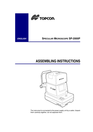
The instrument is connected to the power supply unit by a cable. Unpack them carefully together, do not separate them.
1
Attach the rail covers after removing the locking brackets.
1 Move the instrument body to the right. (seen from the front of the monitor) 2 Remove the two sets of screws securing the locking bracket on the left side of the instrument base. Remove the bracket. 3 Attach and screw the rail cover to the instrument base using the sets of screws removed in step 2. 4 Move the instrument body to the left and remove the right locking bracket. Attach the rail cover. Note:
Keep the locking brackets for future use.
2
Checking the voltage selector Make sure that the voltage selector is set at the proper voltage.
100V 120V
230V 240V
VOLTAGE SELECTOR
Note:
2
If not, contact your authorized TOPCON dealer.
3
Checking the cross-slide operation Manipulate the control lever to make sure the cross-slide moves smoothly.
Note:
4
When first unpacking the instrument, the cross-slide may not move smoothly in the right and left direction. If this is the case, aggressively move the cross-slide right and left and any uncharacteristic roughness in movement will go away.
Attaching the chin rest pad Attach the chin rest pad using the two chin rest pins.
3
ENGLISH
INSTRUCTION MANUAL
SPECULAR MICROSCOPE
SP-2000P
Copyrights and trademarks SP-2000P is a trademark of the TOPCON Corporation. © TOPCON 1999
INTRODUCTION
Thank you very much for purchasing the TOPCON SP-2000P Specular Microscope. This Instruction Manual gives a description of the TOPCON SP-2000P. This includes its main features and basic operation, troubleshooting and the checking, maintenance and cleaning of this instrument. Please read both the sections Displays for safe use on page v and Safety Cautions on page vii carefully before putting this instrument into operation. To get the best use from this instrument, read these instructions carefully and place this manual in a convenient location for future reference.
Specular Microscope SP-2000P
This instrument contains the following features ● Photography of the corneal endothelium and measurement of the
corneal thickness; ● The auto-alignment function ensures quick and easy photography and measurements; ● Simplified cell analysis which makes reference values, such as cell density, easily obtainable.
Precautions
● This Specular Microscope is a piece of precision equipment, which
needs to be used and kept in places under normal life conditions regarding temperature and humidity. Do not expose the instrument to direct sunlight. ● To ensure best use, install the instrument on a level floor, free from any vibration. ● Always check that all cables are plugged in correctly before use. WARNING: For your own safety always make sure the instrument is correctly grounded for compatibility with high currents. Never disable the grounding plug of the power cord. ● Use the power at the rated voltage. ● To obtain clear images and to ensure accurate results, make sure the
monitor is not smeared with fingerprints and/or stains. ● TOPCON is not responsible for any modification caused by
disassembly or adjustments made by unauthorized dealers or persons. ● If any trouble occurs with your instrument or its accessories, first refer to the troubleshooting guide in this manual and carry out the checks listed there. If nothing is found during your check, then contact your authorized dealer or TOPCON to service it. ● Always turn the power source off and place the dust cover on the instrument when it is not in operation.
iv
Introduction
Selecting externally connected equipment The TOPCON SP-2000P complies with the CE marking. Before connecting a personal computer, bar code reader, image recorder, or TV monitor to the TOPCON product, make sure that such external equipment is in compliance with the CE marking.
Displays for safe use Important warnings are placed on the products and inserted in the instruction manuals, in order to encourage the safe use of products and prevent any danger to the operator and others or damage to existing facilities. Please familiarise yourself with the following displays and icons before reading the "Safety Cautions" and the further text.
INSTRUCTION MANUAL
v
Specular Microscope SP-2000P
Meaning of displays
Display
Meaning
Ignoring or disregarding this display may result in serious injury or lead to life-threatening situations. WARNING
Ignoring or disregarding this display may lead to personal injury or severe damage to the instrument or facilities. CAUTION ● Injury potential includes hurt, burn, electric shock, etc. ● Damage to facilities refers to extensive damage to buildings,
equipment and furniture.
Meaning of icons
Icons
Meaning This indicates Mandatory Action. Specific content is expressed with words or an image, located close to the icon. This icon indicates a Hazard Alert (Warning). Specific content is expressed with words or an image, located close to the icon. This indicates Prohibition. Specific content is expressed with words or an image, located close to the icon.
vi
Introduction
Safety Cautions This instruction manual specifies safety precautions necessary to prevent accidents. Always observe these precautions and use the instrument correctly.
WARNING Icons
Prevention item
Page
● An electric shock may cause burns or possible fire. Turn
the main power switch OFF and UNPLUG the power cord before replacing fuses. Replace only with fuses of the correct rating.
37
CAUTION Icons
Prevention item
Page
● To avoid potential injury, hold the instrument in the
proper position.
24
● To avoid potential injury and/or damage to the
instrument, do not drop the instrument.
24
● To avoid potential injury, keep your fingers away from
the chin rest.
38
● To avoid potential injury, do not put your hand or finger
under the measuring head when moving the head up and down.
37
● To avoid potential injury during operation, do not touch
the patient’s eyes or nose with the instrument.
INSTRUCTION MANUAL
37
vii
Specular Microscope SP-2000P
CAUTION Icons
Prevention item
Page
● To avoid potential injury, do not push the instrument to
the patient's side when "TOO CLOSE" is displayed on the screen.
37
● To prevent an electric shock, turn main power switch
OFF and UNPLUG the power cord before replacing the flash bulb.
70
● To avoid potential injury, do not touch the locking hook if
the protective tape is no longer wrapped around the hook.
70
● To prevent shock hazard, do not allow water or other
foreign matter to come into contact with the instrument.
24
● To avoid an electric shock, do not open the instrument.
Refer all servicing to qualified personnel only.
67
Operation and maintenance Purpose This specular microscope is a piece of precision electrical equipment for medical use, which must be used according to a doctor’s instructions.
User maintenance To ensure the safety and performance of the instrument, the maintenance shall be carried out by a trained service technician only, unless otherwise specified in this manual. The following maintenance tasks, however, can be performed by the user. We refer you to the applicable text in this manual, with regard to the maintenance method.
viii
Introduction
Fuse replacement
The primary and secondary fuses for the main body can be replaced by a non-trained service technician. For details, we refer you to the applicable text in this manual.
Flash Bulb replacement
The flash bulb for photography can be replaced by a non-trained technician. For details, we refer you to the applicable text in this manual.
Cleaning of Monitor
The cleaning procedure for the lens and glass sections of the monitor is found on page 69. For details, we refer you to the applicable text in this manual.
TOPCON instruments general statements
● TOPCON shall not take any responsibility for damage due to fire,
earthquakes, actions by third persons and other accidents, or the negligence and misuse by the user or use under unusual conditions. ● TOPCON shall not take any responsibility for damage resulting from the inability to use this equipment, such as a loss of business profit and suspension of business. ● TOPCON shall not take any responsibility for damage caused by operations other than those described in this Instruction Manual. ● The diagnoses that are made are the responsibility of relevant doctors, and TOPCON shall not take any responsibility for the results of such diagnoses.
INSTRUCTION MANUAL
ix
Specular Microscope SP-2000P
Warning indications and positions To ensure safe usage of this equipment, precaution indications are provided. Follow the warning instructions. If any of the following labels are missing, please contact us at the address stated on the back cover.
CAUTION • To avoid potential injury, do not put your hand or finger under the measuring head when moving the head up and down.
CAUTION • To avoid potential injury during operation, do not touch the patient’s eyes or nose with the instrument.
CAUTION • To avoid electric shock, do not open the instrument. Refer all servicing to only qualified personnel.
WARNING • Electrical shock may cause burns or possible fire. Turn the main power switch OFF and UNPLUG the power cord before replacing fuses. Replace only with fuses of the current rating.
x
Contents
INTRODUCTION This instrument contains the following features
iv
Precautions
iv
Selecting externally connected equipment...
Displays for safe use
v
v
Meaning of icons... vi
Safety Cautions
vii
Operation and maintenance
viii
Purpose... viii User maintenance... viii
1
TOPCON instruments general statements
ix
Warning indications and positions
x
COMPONENTS 1.1
Main Body
15
1.2
Control panel
17
1.3
Monitor screen
18
1.3.1
Ocular anterior observation screen... 18
1.3.2
Corneal endothelium observation screen... 18
1.3.3
Photographed result image screen... 19
Specular Microscope SP-2000P
1.3.4
Photographed result image screen with analytic values... 19
1.3.5
Simplified cell count mode screen... 20
1.3.6
Menu screen... 20
1.4
2
Standard accessoires
21
PREPARATIONS BEFORE USE 2.1
Assembly procedure
24
2.1.1
Installing the instrument... 24
2.1.2
Connecting the power cord... 25
2.1.3
Connecting the mouse... 26
2.1.4
Connecting the external input/output terminal... 26
2.2 2.2.1
3
Default settings
28
Reset from the power-saving status... 36
BASIC OPERATION 3.1
Preparation for photography
38
3.1.1
Photographing in auto mode... 38
3.1.2
Photographing in manual mode... 44
3.2
4
Displaying and deleting the photographed results
50
OPERATION BY OBJECTS 4.1
Simplified cell analysis
55
4.2
Output to the IMAGEnet System
59
4.3
Output to an image processing unit
60
4.4
Input/output via RS-232C
61
Output via RS-232C... 61 Input via RS-232C... 63
4.5
5
RS-232C communication specifications
64
MAINTENANCE AND CHECKS 5.1
Daily maintenance
68
5.2
Be consistent
68
5.2.1
Photograph window... 68
5.2.2
Adjusting the monitor screen... 68
5.3
Cleaning the photograph window
69
Wiping off stains... 69
xii
5.4
Replacing the flash bulb
70
5.5
Replacing the fuses
71
Contents
5.6
6
72
REFERENCE MATERIAL 6.1
Optional accessories
75
6.1.1
Motorised adjustable instrument table AIT-20 and tabletop... 75
6.1.2
Specifications... 75
6.1.3
Motorised adjustable instrument table AIT-11 and special tabletop... 76
6.1.4
Specifications... 76
6.2 6.2.1
6.3 6.3.1
7
8
Replacement Parts
Related products
77
IMAGEnet system... 77
Terminology
77
Description of terms... 77
TROUBLESHOOTING 7.1
Self-Check
79
7.2
Troubleshooting table
80
SPECIFICATIONS
85
INSTRUCTION MANUAL
xiii
1
COMPONENTS
1.1 Main Body (2)
(1)
(3)
(4)
(5)
(6)
(8) (7)
Specular Microscope SP-2000P
(1)
Lamp housing cover
(2)
Measuring head
(3)
Monitor
(4)
Photography switch
(5)
Joystick
(6)
Power indicator
(7)
Locking knob (for movement prevention)
(8)
Control panel (9) (10)
(11) (15)
(12)
(13)
(17)
(14)
(18) (16)
16
(9)
Forehead rest
(10)
Photography window
(11)
Vertical positioning mark
(12)
Chinrest vertical movement handle
(13)
Chinrest tissue with locking pins
(14)
Cinrest
(15)
Power switch
(16)
Power cord
(17)
Mouse
(18)
Monitor adjusting knobs
Components
1.2 Control panel
SELECT
MODE
FLASH
(21) (19) (22) DELETE
MENU
CLEAR
(23)
(24)
(20) POWER
(19)
Select switch Switches between the display of the photographed result and the ocular anterior observation screens.
(20)
Menu switch Displays the menu screen.
(21)
Flash switch Sets the HIGH/LOW level regarding flash intensity.
(22)
Mode switch Sets the AUTO/MANUAL mode of image capturing.
(23)
Clear swicht Deletes all photographic results stored in the memory.
(24)
Delete switch Deletes photographic results being stored in the memory.
INSTRUCTION MANUAL
17
Specular Microscope SP-2000P
1.3 Monitor screen 1.3.1
Ocular anterior observation screen
AUTO
LOW
L0001
(25)
(27)
(26)
(28) (29)
S N C T I (25)
Image capturing mode
(26)
Intensity flash
(27)
Photograph serial No.
(28)
Position indication eye
(29)
Alignment circle
(30)
Photography point switching icon
1.3.2
(30)
Corneal endothelium observation screen
Longitudial alignment bar
18