VERATHON Inc
AMI 9700 Measurement Quick Reference
Quick Reference
4 Pages

Preview
Page 1
AMI 9700 Measurement Quick Reference
A Critical Measure of Patient Health
Turn on the AMI 9700. Turn on the instrument by pressing the POWER ON/OFF button.
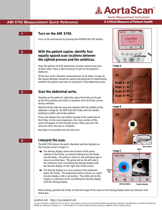
With the patient supine, identify four equally spaced scan locations between the xiphoid process and the umbilicus. Image A
Have the patient lie with abdominal muscles relaxed and arms at their sides. Place a liberal amount of gel on the patient’s abdomen. Of the four aortic diameter measurements to be taken (Image A), the largest diameter should be used by the physician for determining whether the patient may have an aneurysm in the abdominal aorta.
Xiphoid Process
Scan 1 Scan 2 Scan 3 Scan 4
Scan the abdominal aorta. Standing at the patient’s right side, place the Probe on the gel at the first position and hold it in position with the Probe screen facing vertically.
Umbilicus
Image B
Hold the Probe with the long axis aligned with the midline of the abdomen (Image B). Do NOT hold the Probe with the handle pointing to either side of the patient. Press and release the scan button located at the underside of the Probe. As the scan progresses, the cross-section of the aorta will appear on the Console screen. When you hear the end-scan tone, the scan is complete. See step 6 to annotate and save the scan.
Interpret the scan. The AMI 9700 shows the aortic diameter and two displays on the Console screen (Image C):
Scan button
Image C
The Aiming display shows the location of the aorta relative to the Probe, as viewed looking from the Probe into the body. The aorta is shown in red and bowel gas is shown as white lines. The green dot on the left side is the reference mark correlating the Aiming display with the Results display on the right side of the screen. The Results display is a cross-section of the abdomen below the Probe. The abdominal aorta is shown as a dark circular shadow with a red outline. The white dot on the image is a reference mark correlating the Results display with the Aiming display.
Aorta
Bowel gas
Aiming display
Results display
When aiming, position the Probe so that the image of the aorta on the Aiming display does not intersect with bowel gas.
verathon.com https://my.scanpoint.com For more information, call 800.331.2313 (in the U.S. and Canada) or contact your local Verathon Medical representative. AortaScan, NeuralHarmonics, ScanPoint, Verathon and VMODE are trademarks of Verathon Inc. © 2009 Verathon Inc. All other brands and product names are trademarks of their respective owners.
AMI 9700 9700 Measurement Measurement Quick Quick Reference Reference AMI
(cont.)
A Critical Measure of Patient Health
Interpreting the Scan - Conditions Affecting Scan Accuracy Measuring Aortic Diameters < 3 cm (Image D) The AMI 9700 can detect aortas with diameters between 3 cm and 12.4 cm. Aortas with diameters less than 3 cm will occur in patients who have normally-sized aortas.
Image D
The round shadow at 6 cm depth in the Results display is the patient’s abdominal aorta. Patients with aortas less than 3 cm in diameter will show no red outline around the aorta in the Results display. With aortas less than 3 cm in diameter, the diameter cannot be measured automatically, but a measurement can be made using Manual Measurement Mode (step 5).
Partial Gas Obstruction (Image E) A green arrow on the Console and a solid green arrow on the Probe indicate the abdominal aorta can be detected, but the presence of bowel gas prevents an accurate measurement.
Image E
Moving the Probe 1/2 to 1 inch (1 to 2 cm) in the direction of the arrow has a high probability of providing a successful scan. In this case, the Probe should be repositioned and the patient rescanned. Gently but firmly work the probe into the tissues of the abdomen with a side-to-side rocking motion to try and displace any bowel gas obscuring the aorta.
Substantial Gas Obstruction (Image F) A red arrow on the Console and a flashing green arrow on the Probe indicate bowel gas has substantially obscured the aorta. No diameter measurement can be calculated. Although moving the Probe 1/2 to 1 inch (1 to 2 cm) in the direction of the arrow has a low probability of providing a successful scan, an additional scan should be attempted. In this case, the Probe should be repositioned and the patient rescanned. Gently but firmly work the probe into the tissues of the abdomen with a side-to-side rocking motion to try and displace any bowel gas obscuring the aorta. If rescanning is not successful, the exam should be postponed and rescheduled. Have the patient fast for 12 hours prior to exam.
verathon.com https://my.scanpoint.com
Image F
BVI Mode Quick Reference AMI 9700 Measurement Quick Reference AMI9600 9700AortaScan Measurement Quick Reference TM
(cont.)
A Critical Measure of Patient Health
Interpreting the Scan - Conditions Affecting Scan Accuracy Obesity Attenuation of the ultrasound signal by excess abdominal fat can result in a poor ultrasound image, which affects the quality of the diameter measurement. With obese patients, try pressing the Probe firmly into the abdomen to reduce the distance to the aorta as much as possible, while attempting to minimize patient discomfort. In rare cases, it is possible for a patient’s abdomen to be too thick for the ultrasound to reach the aorta. The depth of the ultrasound beam is 18 cm. If a patient has an extra thick abdomen where the distance from the Probe face to the aorta is greater than 18 cm, the AMI 9700 will not detect the aorta. In these cases, other imaging methods should be used.
Manual Measurement of Aortic Diameter
Image G
Should the user desire, manual measurement of the diameter of the aorta is permitted: a. After completing the scan, activate Manual Measurement Mode . b. Use the Axis Select icon to select either the “Up-Down” or “Left-Right” axis of movement for the highlighted cursor (Image G). c. Move the highlighted cursor to the edge of the aortic cross-section shown in the Results display. d. Select the other cursor using the Cursor Select icon (Image H). e. Move the second cursor on to the opposite edge of the aortic cross-section in the Results display. f.
Axis select
Image H
Press the Return icon to exit Manual Measurement Mode (Image H).
Cursor select
verathon.com https://my.scanpoint.com
Return
AMI 9700 Measurement Quick Reference
Save, review, and print exam results.
A Critical Measure of Patient Health
Image I
To save the exam, you must provide a voice annotation. The AMI 9700 does NOT automatically save each scan. It is recommended that the user adds a voice annotation or writes down the diameter calculated for each location. To annotate, press the RECORD button on the Console. When you see the RECORD button icon change to a STOP button icon, record the patient information by speaking into the Probe microphone. Press STOP when finished. When the hourglass icon disappears, press the LISTEN button to replay the annotation (Image I). To print exam results via the onboard printer, press the PRINT button. To view prior exam results, press the REVIEW button. On the Review Screen, two types of diameter measurements may be displayed (Image J):
Image J
DiameterV-mode – Diameter measured automatically by AMI 9700. DiameterManual – Diameter measured manually in Manual Measurement Mode. DiameterManual will only be displayed if a manual measurement was made. To perform another exam, press the HOME button.
Scan the aorta at the remaining 3 locations. Repeat steps 3, 4 and 6 at three more locations spaced at equal distance between the xiphoid process and the umbilicus. The largest aortic diameter measured should be used by the physician to help determine if the patient has an aneurysm in the abdominal aorta.
Finish the exam Once you have completed the exam, wipe the ultrasound gel off the patient and Probe. For ScanPointTM subscribers, logging onto the ScanPointTM application automatically transfers and saves your annotated exams to your Windows® computer.
To order additional rolls of paper (0800-0319) or batteries (0400-0066), contact Customer Care at 800.331.2313.
verathon.com https://my.scanpoint.com
0123
For more information, call 800.331.2313 (in the U.S. and Canada) or contact your local Verathon Medical representative. Verathon Corporate Headquarters: 20001 North Creek Parkway, Bothell, WA 98011, USA. Phone: +1.425.867.1348 VM Europe B.V.: Linnaeusweg 11, 3401 MS IJsselstein, The Netherlands, Phone:+31.30.68.70.570
0900-2198-03-60