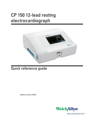
Quick Reference Guide
16 Pages

Preview
Page 1
CP 150 12-lead resting electrocardiograph
Quick reference guide
© 2013 Welch Allyn, Inc. To support the intended use of the product described in this publication, the purchaser of the product is permitted to copy this publication, for internal distribution only, from the media provided by Welch Allyn. Caution: Federal US law restricts sale of the device identified in this manual to, or on the order of, a licensed physician. Welch Allyn assumes no responsibility for any injury, or for any illegal or improper use of the product, that may result from failure to use this product in accordance with the instructions, cautions, warnings, or indications for use published in this manual. Welch Allyn is a registered trademark of Welch Allyn, Inc. CP 150, and CardioPerfect are trademarks of Welch Allyn, Inc. Welch Allyn Technical Support: http://www.welchallyn.com/about/company/locations.htm 105755 (CD) DIR 80018918 Ver. A
Material Number 721331, DIR 80018918 Ver. A
Regulatory Affairs Representative Welch Allyn 4341 State Street Road
Welch Allyn Limited
Skaneateles Falls, NY 13153-0220 U.S.A
Dublin Road
www.welchallyn.com
Navan Business Park Navan, County Meath Republic of Ireland
1
Powering the electrocardiograph The electrocardiograph runs on AC or battery power. Plug the electrocardiograph into AC power as often as possible so that the built-in charger can keep the battery charged. Regardless of the battery condition, you can use the electrocardiograph whenever it is plugged in. WARNING When you use AC power, always plug the electrocardiograph into a hospital-grade outlet to avoid the risk of shock. WARNING If the integrity of the building’s safety ground is in doubt, operate this device on battery power to avoid the risk of shock. To turn on or turn off
Press
.
Connecting the patient cable
2
Powering the electrocardiograph
CP 150 12-lead resting electrocardiograph
WARNING Do not allow the conductive parts of the patient cable, electrodes, or associated connections of defibrillation-proof applied parts, including the neutral conductor of the patient cable and electrodes, to come into contact with other conductive parts, including earth ground. Otherwise, an electrical short might result, risking electric shock to patients and damage to the device. WARNING To avoid injury to the patient or damage to the device, never plug patient leads into any other device or wall outlet. WARNING To provide CF protection use only accessories approved by Welch Allyn. Visit www.welchallyn.com. The use of any other accessories can result in inaccurate patient data, can damage the equipment, and can void your product warranty. CAUTION Always connect the patient cable and the leads properly during defibrillation. Otherwise, the connected leads could be damaged.
Loading the thermal paper The electrocardiograph prints on z-fold thermal paper. •
Store the paper in a cool, dry, dark place.
•
Do not expose it to bright light or UV sources.
•
Do not expose it to solvents, adhesives, or cleaning fluids.
•
Do not store it with vinyls, plastics, or shrink wraps.
3
Attaching the leads to the patient Proper lead attachment is important for a successful ECG. The most common ECG problems are caused by poor electrode contact and loose leads. Follow your local procedures for attaching the leads to the patient. Here are some common guidelines. WARNING Electrodes can cause allergic reactions. To avoid this, follow the electrode manufacturer’s directions. To attach the leads to the patient 1. Prepare the patient. •
Describe the procedure. Explain the importance of holding still during the test. (Movement can create artifact.)
•
Verify that the patient is comfortable, warm, and relaxed. (Shivering can create artifact.)
•
Put the patient in a reclining position with the head slightly higher than the heart and legs (the semi-Fowler’s position).
2. Select the electrode locations. (See the “Electrode locations” chart.) •
Look for flat areas.
•
Avoid fatty areas, bony areas, and major muscles.
3. Prepare the electrode locations. •
Shave or clip the hair.
•
Thoroughly clean the skin, and lightly rub it dry. You may use soap and water, isopropyl alcohol, or skin preparation pads.
4. Attach the lead wires to the electrodes. 5. Apply the electrodes to the patient.
4
Attaching the leads to the patient
CP 150 12-lead resting electrocardiograph
Electrode examples, left to right: arm clamp (reusable), Welsh cup (reusable), tab electrode (disposable), monitoring electrode (disposable). •
For reusable electrodes: Use electrode paste, gel, or cream to cover an area the size of each electrode but no larger. Secure the arm and leg clamps. Apply the Welsh cups (suction electrodes) to the chest.
•
For disposable tab electrodes: Place the electrode tab between the “jaws” of the connector. Keep the tab flat. Verify that the metal portion of the connector makes contact with the skin side of the electrode tab.
•
For all disposable electrodes: Lightly tug on the connector to ensure that the lead is securely attached. If the electrode comes off, replace it with a new electrode. If the connector comes off, reconnect it.
5
ECG home screen ECG home screen The ECG home screen includes the following areas:
Item
Area
1
Device status
2
Content
3
Navigation
Device status area The Device status area, located at the top of the ECG home screen, displays: •
Time and date
•
Battery status
•
Error or information messages. These items are displayed until the condition has been resolved.
6
ECG home screen
CP 150 12-lead resting electrocardiograph
Content area The Content area includes 3 test selection buttons and a preview selection button: •
Auto ECG
•
Rhythm ECG
•
Stat ECG
•
Electrode Placement
The content area also provides shortcuts to several controls.
About the test types Auto ECG
A report typically showing a 10-second acquisition of 12 leads of ECG information combined with patient data, measurements, and optional interpretation. Auto ECGs can be saved to the electrocardiograph’s test directory or to a USB mass-storage device.
Rhythm ECG
A continuous, real-time printout of rhythm strips with a user-defined lead configuration. Rhythm ECGs are printouts only. They cannot be saved.
Stat ECG
An auto ECG that starts without waiting for you to enter patient data.
Navigation area The Navigation area includes the following tabs: •
ECG home: Displays ECG test types and provides shortcuts to several controls.
•
Manage worklist : Includes patient data entered manually or orders downloaded when connected to a hospital information system.
•
Saved tests: Accesses the patient ECG tests.
•
Settings: Accesses device configuration settings.
To navigate to a tab, touch the tab in the Navigation area with the corresponding name. The active tab is highlighted.
7
Performing an Auto ECG test CAUTION Patient data is not saved until the ECG test is completed. The ECG configuration settings can be changed in the Settings tab. The following settings may appear differently if the default settings have been modified.
Note
1. Touch the
(Auto ECG) button. The Summary tab appears.
2. Enter the following patient information as desired: •
Patient ID. Touch the OK button.
•
Birth date. Touch the OK button.
•
Last name. Touch the OK button.
•
First name. Touch the OK button.
•
Middle Initial. Touch the OK button.
Note 3. Touch the
If the patient has a pacemaker touch the Pacemaker present button. (Next) button.
4. Enter the following patient information as desired: •
Gender
•
Race
•
Height. Touch the OK button.
•
Weight. Touch the OK button.
•
Comments. Touch the OK button.
5. Touch the
(Next) button.
6. Attach the leads to the patient. Note
If desired, touch the (torso) button to enlarge the lead-placement screen. Any flashing dots on the ECG preview screen indicate unattached or poorly attached leads.
8
Performing an Auto ECG test
CP 150 12-lead resting electrocardiograph
Item
Button
1
Leads button
2
ECG preview button
3
Gain button (size)
4
Speed button
5
Filters button
7. If an Artifact message appears, minimize the artifact, as described under Troubleshooting. You might need to ensure that the patient is comfortably warm, reprepare the patient’s skin, use fresh electrodes, or minimize patient motion. 8. (Optional) Adjust the waveforms, using the buttons to cycle through the following options: •
leads displayed
•
ECG preview format
•
gain (size)
•
speed
•
filters
9. Touch the Print button to perform the Auto ECG test. If Print preview on is selected in the ECG configuration settings, a single screen of the ECG Report Print Preview appears. Touch Print to continue with the Auto ECG test or touch Retest to discard the test and return to the previous screen. 10. If a Waiting for 10 seconds of quality data message appears, at least 10 seconds of ECG data have been collected with excessive artifact. Time requirements in the message may vary, based upon selected print format. Minimize the artifact, as described under Troubleshooting. Then wait for the test to print. If necessary, you can override the wait time and print the available data immediately, but the result might be an incomplete or poor-quality test.
Quick reference guide
Performing an Auto ECG test
9
11. After the test prints, select the desired option: Reprint, Save, Retest, or Rhythm. If the Auto Save setting is turned off, touch Save to save the test. Select one of the following locations: •
Local (internal memory)
•
USB mass storage device (Any tests that you save to a USB mass storage device can be retrieved only from a CardioPerfect workstation.)
•
Workstation
•
Remote file location
12. Touch Reprint to print the test again, touch Retest to discard the test and start over, or touch Exit. WARNING To avoid the risk of associating reports with the wrong patients, make sure that each test identifies the patient. If any report does not identify the patient, write the patient identification information on the report immediately following the ECG test.
11
Troubleshooting Lead-quality problems “Artifact” message on the screen Artifact is signal distortion that makes it difficult to accurately discern the waveform morphology. Causes •
The patient was moving.
•
The patient was shivering.
•
There is electrical interference.
Actions See actions for wandering baseline, muscle tremor, and AC interference.
Wandering baseline Wandering baseline is an upward and downward fluctuation of the waveforms.
Causes •
Electrodes are dirty, corroded, loose, or positioned on bony areas.
•
The electrode gel is insufficient or dried.
•
The patient has oily skin or used body lotions.
•
Rising and falling of chest during rapid or apprehensive breathing.
Actions •
Clean the patient’s skin with alcohol or acetone.
•
Reposition or replace the electrodes.
•
Verify that the patient is comfortable, warm, and relaxed.
•
If wandering baseline persists, turn the baseline filter on.
12
Troubleshooting
CP 150 12-lead resting electrocardiograph
Muscle tremor
Causes •
The patient is uncomfortable, tense, nervous.
•
The patient is cold and shivering.
•
The exam bed is too narrow or short to comfortably support arms and legs.
•
The arm or leg electrode straps are too tight.
Actions •
Verify that the patient is comfortable, warm, and relaxed.
•
Check all electrode contacts.
•
If interference persists, turn the muscle-tremor filter on. If interference still persists, the problem is probably electrical in nature. See the suggestions for reducing AC interference (in a related troubleshooting tip).
AC interference AC interference superimposes even-peaked, regular voltage on the waveforms.
Causes •
The patient or technician was touching an electrode during recording.
•
The patient was touching a metal part of an exam table or bed.
•
A lead wire, patient cable, or power cord are broken.
•
Electrical devices in the immediate area, or lighting, or wiring concealed in walls or floors are interfering.
•
An electrical outlet is improperly grounded.
•
The AC filter is turned off or set incorrectly.
Actions •
Verify that the patient is not touching any metal.
•
Verify that the AC power cable is not touching the patient cable.
•
Verify that the proper AC filter is selected.
•
If interference persists, unplug the electrocardiograph from AC power and run it on the battery. If this solves the problem, you’ll know that the noise was introduced through the power line.
•
If interference still persists, the noise may be caused by other equipment in the room or by poorly grounded power lines. Try moving to another room.
Quick reference guide
Troubleshooting
13
Lead alert or square wave A dot might be flashing on the lead-status screen. Or one or more leads might appear as a square wave. Causes •
Electrode contact might be poor.
•
A lead might be loose.
•
A lead might be defective.
Actions •
Replace the electrode.
•
Verify that the patient’s skin has been properly prepared.
•
Verify that electrodes have been properly stored and handled.
•
Replace the patient cable.
Material No.
721331