User Guide
72 Pages

Preview
Page 1
• Streak Retin Foreign2
11/10/98
4:58 PM
Page 2
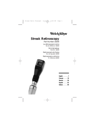
Thank you for purchasing the Welch Allyn No. 18200 3.5v halogen streak retinoscope. This instrument has been designed to meet the needs of today’s practitioners and incorporates features not found on any other retinoscope: 1. External Focusing Sleeve-unique planetary gear system allows for easy adjustment no matter what size hand or how instrument is held. Continuous 360° rotation. Maintains the same plane of focus during rotation. 2. Improved Light Output-brighter halogen lamp provides 50 percent more intensity than previous lamps. The reflex is now crisper and easier to see in all patient’s eyes. Retinoscopy can be done faster and more accurately. 3. Dust-free Optics-new housings and glass cover on the front keep the instrument cleaner longer. 4. Crossed Linear Polarizing Filter-dramatically reduces glare from lenses. Allows retinoscopy to be performed closer to the axis of the correcting lenses. 5. Fixation Cards-new cards that easily attach increase the ease with which dynamic retinoscopy is performed. 6. Improved Optics-glare and shadows have been eliminated for a clearer and more precise view. 7. Interchangeability-By simply changing the lamp, the streak retinoscope can be converted to a spot retinoscope.
ii
• Streak Retin Foreign2
11/10/98
4:58 PM
Page 1
Introduction Retinoscopy is a technique for objective refraction of the eye. There are two basic types of retinoscopy. Static retinoscopy (described in this booklet) is done with the patient fixating at a distance. Dynamic retinoscopy is done with the patient fixating at a near target. These techniques require diligence and expertise that can result in a precise measurement of the refractive error of an eye. There are two types of self-illuminated retinoscopes. The streak retinoscope featured in this booklet is the most widely used scope today and has largely supplanted the spot retinoscope. In retinoscopy, a parallel or slightly divergent beam of light is directed into the patient’s eye. This results in illumination of the retina, and the reflected light from the retina causes reflexes observed by the examiner in the patient’s pupil. The refractive status of the eye is found by using correcting trial lenses to make the far point of the ametropic eye conjugate to the pupil of the examiner’s eye. When this is achieved, the movement of the reflex will be neutralized. The material in the booklet is presented with the assumption that the reader is familiar with retinoscopy in general. Two excellent references for retinoscopic technique are: Corboy, J.M.; The Retinoscopy Book, 3rd Edition, 1989, SLACK Incorporated. or on videotape: Guyton, D.L.; Retinoscopy: Minus Cylinder Technique or Retinoscopy: Plus Cylinder Technique, 1986, American Academy of Ophthalmology-Continuing Ophthalmic Video Education.
1
• Streak Retin Foreign2
11/10/98
4:58 PM
Page 2
The streak retinoscope is found by most practitioners to be easy to use, fast, accurate, and especially valuable in determining the axis of astigmatism. There are several features of the streak retinoscope that make determining the refractive state of the eye easy and accurate. These are: 1. Each meridian can be neutralized separately. 2. All errors can be neutralized using either “with” or “against” motion or perhaps using both. 3. The axis of astigmatism is apparent. 4. Streak retinoscopy is easy because one watches a band of light instead of a shadow. 5. Streak retinoscopy may easily be done with undilated pupils.
2
• Streak Retin Foreign2
11/10/98
4:58 PM
Technique 1. The Operation of the Control Sleeve of the Scope The operator will note that the width of the streak varies as the sleeve is raised and lowered (see Figure 1). When the operating sleeve is in the lowest position the light rays emitted are slightly divergent. Here the instrument acts with a plano mirror effect, which reflects divergent rays that will never come to a focus. As the sleeve is raised, the streak focuses. With the sleeve all the way up, the retinoscope acts with a concave mirror effect, where the light rays cross and then diverge. Because the rays cross, the eye’s reflex moves in opposite directions with the concave mirror effect as compared to the plano mirror effect. Throughout this booklet, we will use the plano mirror effect unless specified. The rotary movement of the control sleeve mechanism allows the streak to rotate 360° to ascertain the axis of astigmatism (see Figure 1). 2. Preliminary Steps A) Set the sleeve in its lowest position (plano-mirror effect).
Figure 1
3
Page 3
• Streak Retin Foreign2
11/10/98
4:58 PM
Page 4
B) Position yourself 2/3 meter (26") from the patient. This distance implies a working lens of +1.50D (computed as the reciprocal of working distance in meters). Working distance and lens may be varied to suit the practitioner’s needs. (In this instruction book the 2/3 meter (26") working distance is assumed. Different working distances can be used, but remember to adjust for your working distance.) C) With the refracting equipment in place, direct the patient’s attention to a fixation spot at 15 feet or more from the eye and align the streak vertically.
Figure 2 No oblique astigmatism
Figure 3 Oblique astigmatism
D) Observe the “reflex” which will appear as in Figure 2, providing no oblique astigmatism is present. If oblique astigmatism is present, the reflex will appear more like Figure 3, where the reflex does not appear vertical. E) Move the vertical streak horizontally across the pupil and back again and observe whether the reflex moves in the same direction as the streak or in the opposite direction.
4
• Streak Retin Foreign2
11/10/98
4:58 PM
Page 5
F) Rotate the control sleeve until the streak is horizontal and move the streak vertically. The reflex will appear as in Figures 4 or 5.
Figure 4 No oblique astigmatism
Figure 5 Oblique astigmatism
G) If the streak and the reflex move in the same direction with no lens in the refractive apparatus, the refraction is one of these: 1. Hyperopia; 2. Emmetropia; 3. Myopia of less than 1.50 diopters. If the reflex moves in the opposite direction, the error is myopia greater than 1.50 diopters. 3. Determining Refractive Error By Neutralization Before starting, make sure the eye not being refracted has some “against” motion using the plano mirror effect. This will blur vision to prevent accommodation. If “with” or neutral motion is noticed initially, place about a +1.00 sphere before the eye once neutral motion is seen. A) Neutralizing with spheres only: 1. Change sphere in the minus direction until the reflexes in all axes have “with” motion. 2. Adjust in the plus direction until the reflex fills the pupil in one meridian and all motion has stopped. This will be one of the principal meridians if astigmatism is present. That meridian is then said to be neutralized.
5
• Streak Retin Foreign2
11/10/98
4:58 PM
Page 6
3. Test for neutralization by one of these methods: a) Move the sleeve all the way up (concave mirror position); the reflex should also appear neutralized; b) Move closer to the patient and “with” motion should return; move away and “against” motion should appear, or c) Place an extra +0.25 sphere in the apparatus and “against” motion should appear; 4. Repeat the neutralization in the meridian 90° away. B) Locating the axis of astigmatism: Two phenomena help in determining the axis of astigmatism: break and width. Break is observed when the streak is not aligned with a principal meridian of the astigmatism (Figures 3 and 5). The streak will be aligned with a principal meridian when the break effect disappears and the width of the reflex is narrowest (and it appears its brightest) (Figure 6).
Figure 6
Proceed with neutralization as before-neutralizing one principal meridian first, then 90° away to neutralize the second principal meridian (Figures 7 and 8).
Figure 7
Figure 8
6
• Streak Retin Foreign2
11/10/98
4:58 PM
Page 7
4. Interpretation of Results A) Hyperopia 1. Hyperopia exists when, at the 2/3 meter distance using the plano mirror effect, “with” motion is neutralized using a plus lens greater than +1.50 diopters and both meridians neutralize with the same strength lens. 2. Total hyperopia is estimated by subtracting 1.50 diopters from the total strength lens used. For example, if it takes a +2.50 lens to neutralize motion at 2/3 meter, the total hyperopic error is +1.00 diopter. B) Myopia 1. Myopia exists under several circumstances. a) When “with” motion, using the plano mirror effect at 2/3 meter, is neutralized with a plus lens of less than 1.50 diopter strength. (When motion is neutralized with exactly a 1.50 diopter lens, the eye is emmetropic.) b) When at 2/3 meter, using the plano mirror effect, no motion appears at all. In other words, when the motion is neutralized with no lens in the refracting apparatus. The myopia is then exactly 1.50 diopters. c) When the motion is “against” using the plano mirror effect, and is neutralized with a minus lens. C) Astigmatism 1. Astigmatism exists when the two principal meridians neutralize with different strength lenses. It may be present in many forms. a) Simple hyperopic; b) Simple myopic; c) Compound hyperopic; d) Compound myopic; e) In the mixed form (one meridian hyperopic and the opposite one myopic).
7
• Streak Retin Foreign2
11/10/98
4:58 PM
Page 8
2. Astigmatism can be measured in one of two ways: a) Neutralize one principal meridian first. Then add the appropriate plus or minus cylindrical lens until the other principal meridian is neutralized. b) Neutralization may also be done by continuing to add spherical lenses until the second principal meridian is neutralized. Then the astigmatic error is equal to the difference in strength of lenses necessary to neutralize the two meridians. 5. Special Considerations A) Axis of astigmatism: Extreme care must be used in setting the axis of the cylinder. If the correcting cylinder is of the proper power, a 10° error in axis will produce a new astigmatism of approximately one third of the strength of the original astigmatism with its principal meridian at approximately 45° to those of the original astigmatism. The technique for setting the axis is referred to as “straddling”. When you have an approximate correction of the refractive error and wish to refine the axis setting, the following technique will be helpful. Move up closer to the eye so that the edges of the reflex can be seen, and compare the widths of the two reflexes as you rotate the streak 45° to either side of the correcting cylinder axis. Recede slowly while doing this. Compare the widths of the two reflexes. If there is an axis error, the reflex will be different widths in the two positions. If you are using plus cylinders, rotate the axis toward the narrow band until the reflex widths are equal. With minus cylinders, move the axis away from the narrow band. When the reflex widths are equal, the proper axis has been determined. It is important that the spherical and cylindrical strength be checked again after completion of this maneuver.
8
• Streak Retin Foreign2
11/10/98
4:58 PM
Page 9
Other Features This retinoscope was designed with the needs of today’s practitioner in mind. Listed below are several additional features that will increase your diagnostic capability. DYNAMIC RETINOSCOPY-The Welch Allyn 3.5v Halogen streak retinoscope can be outfitted with magnetic fixation cards (No. 18250) to help perform dynamic retinoscopy. In dynamic retinoscopy, the patient is asked to fixate on words, shapes or another age-appropriate target in the plane of or even on the retinoscope itself. Dynamic retinoscopy is usually done immediately after completing static retinoscopy. There are many methods of dynamic retinoscopy, among these are book retinoscopy, bell retinoscopy, MEM (Monocular Estimation Method) retinoscopy and near retinoscopy.
Some uses of Dynamic Retinoscopy: 1. To check for accommodative disorders; 2. To obtain refractive information;
9
• Streak Retin Foreign2
11/10/98
4:58 PM
Page 10
3. To help decide amplyopic therapy; 4. To determine the adequacy of cyclopegia. For more information on Dynamic Retinoscopy, read the following: Guyton, D.L., O’Connor, G.M.: Dynamic Retinoscopy, Current Opinion in Ophthalmology 1991, 2:78-80. Locke, L.C., Somers, W.: A Comparison Study of Dynamic Retinoscopy Techniques. Optom Vis Sci 1989, 66:540-544. Mazow, M.L., France, T.D., Finkleman, S., Frank, J.: Acute Accommodative and Convergence Insufficiency. Trans Am Ophthalmol Soc. 1989, 87:158-173.
SPIRALLING-Spiralling is a method for estimating ametropia without lenses. This Figure 9 can be helpful in determining the starting point for lens introduction when working with patients who have a high unknown ametropia. The Welch Allyn No. 18200 streak retinoscope is particularly well suited for this technique because the instrument maintains the same focal plane during streak rotation. CROSSED LINEAR POLARIZING FILTER-A crossed linear polarizing filter can be engaged by moving the sliding switch on the practitioner’s side of the instrument from the down to the up position (see Figure 9). This filter cuts down on reflections and allows retinoscopy to be performed closer to the axis of the correcting lens. SPOT RETINOSCOPY-The Welch Allyn No. 18200 streak retinoscope can be converted to a spot retinoscope by simply changing the lamp. In recent years the spot retinoscope has been largely supplanted by the streak but there are some practitioners who still favor the spot retinoscope.
10
• Streak Retin Foreign2
11/10/98
4:58 PM
Page 11
Some arguments presented for using the spot retinoscope are: 1. When working with pediatric patients, it is important to get the most information in the shortest amount of time. The spot scope, based on the shape of the reflex, can help detect astigmatism very quickly. Also significant amounts of myopia and hyperopia can be determined rapidly. 2. For vision screening a large number of patients, e.g. school screenings, the spot retinoscope can help provide more information in a shorter period of time. 3. For judging the fit of hard contact lenses the spot scope can be helpful in assessing power correction, centration, tear film layer, etc.; and to check soft lenses for indications of buckling, lens transparency, and steep or flat corneal correspondence. To convert the No. 18200 streak retinoscope to a spot retinoscope, simply insert the No. 08300 lamp (see directions on page 12). The spot retinoscope is used in the plano mirror position (sleeve all the way down). For more information on Spot Retinoscopy, read the following: Borish, I.M.; Clinical Refraction 3rd Edition, 1970, The Professional Press Inc. Pg 672.
Cleaning Instructions Retinoscope-External housings may be cleaned with a mild detergent and soft cloth. Do not immerse. Windows may be cleaned with alcohol on a cotton swab or lens paper. Fixation Cards-May be cleaned with a mild detergent.
11
• Streak Retin Foreign2
11/10/98
4:58 PM
Page 12
Instructions for Lamp Replacement No. 18200 Streak Retinoscope (Use only Welch Allyn 3.5v Halogen Lamp No. 08200) No. 18300 Spot Retinoscope (Use only Welch Allyn 3.5v Halogen Lamp No. 08300) 1. Remove retinoscope from power source.
2. Remove lamp: Lift out with nail file, letter opener or similar instrument under base flange.
CAUTION: Lamp may be hot and should be allowed to cool before removal.
12
• Streak Retin Foreign2
11/10/98
4:58 PM
Page 13
3. Insert new lamp: 08200 lamp-line up pin on lamp with slot between the metal electrical contact wires. Push lamp into receptacle as far as it will go. Slot
Electrical contact wires
08300 lamp-push lamp into receptacle as far as it will go. Lamp should insert easily-do not force. Lamp base contact pin should be even with metal cutouts in retinoscope base.
4. Replace retinoscope on power source. The No. 18200 and No. 18300 are essentially the same instrument. The No. 18200 streak retinoscope can be converted to a spot retinoscope by simply inserting the 08300 lamp, and vice versa.
13
• Streak Retin Foreign2
11/10/98
5:00 PM
Page 70
We wish to express our sincere appreciation to Al Fors, O.D., Southern College of Optometry, Memphis, TN; David L. Guyton, M.D., The Wilmer Institute at the Johns Hopkins Hospital, Baltimore, MD; Neil Hodur, O.D., Illinois College of Optometry, Chicago, IL; and John Hoepner, M.D. and Mary Jackowski, O.D., SUNY Health Science Center, Syracuse, NY for their valuable assistance in the preparation of this instructional book. Nous souhaitons exprimer notre sincère appréciation à Al Fors, O.D. (Southern College of Optometry, Memphis, TN), David L. Guyton, M.D. (Wilmer Institute, John Hopkins Hospital, Baltimore, MD), Neil Hodur, O.D. (Illinois College of Optometry, Chicago, IL), John Hoepner, M.D, et Mary Jackowski, O.D. (SUNY Health Science Center, Syracuse, NY), pour leur précieuse contribution à la préparation de cette brochure. Den folgenden Personen möchten wir herzlich für ihre wertvolle Unterstützung bei der Ausarbeitung dieser Broschüre danken: Al Fors, O.D., Southern College of Optometry, Memphis, TN; David L. Guyton, M.D., The Wilmer Institute am Johns Hopkins Hospital, Baltimore, MD; Neil Hodur, O.D., Illinois College of Optometry, Chicago, IL; sowie John Hoepner, M.D. und Mary Jackowski, O.D., SUNY Health Science Center, Syracuse, NY. Deseamos expresar nuestros sinceros agradecimientos a los Dres. Al Fors, O.D. Southern College of Optometry, Memphis, Tennessee; David L. Guyton, M.D., The Wilmer Institute del Johns Hopkins Hospital, Baltimore, Maryland; Neil Hodur, O.D., Illinois College of Optometry, Chicago, Illinois y John Hoepner y la Dra. Mary Jackowski, O.D., SUNY Health Science Center, Syracuse, NY, por su valiosa asistencia en la preparación de este libro de instrucciones. Desideriamo esprimere la nostra sincera gratitudine ad Al Fors, O.D., Southern College of Optometry, Memphis, TN; David L. Guyton, M.D., The Wilmer Institute at the Johns Hopkins Hospital, Baltimore, M.D.; Neil Hodur, O.D., Illinois College of Optometry, Chicago, IL; e John Hoepner, M.D. e Mary Jackowski, O.D., SUNY Health Science Center, Syracuse, NY per il prezioso apporto alla stesura di questo manualetto.
Welch Allyn, Inc. 4341 State Street Road, P.O. Box 220 Skaneateles Falls, NY 13153-0220 U.S.A. Phone: (315) 685-4560 FAX: (315) 685-3361 Part No. 182034ML Rev. B
Printed in U.S.A.